5A0E

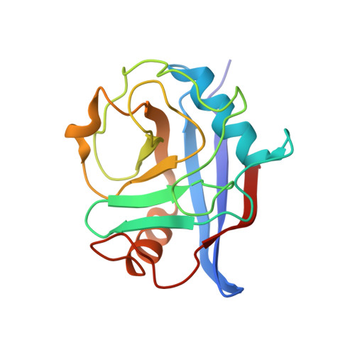
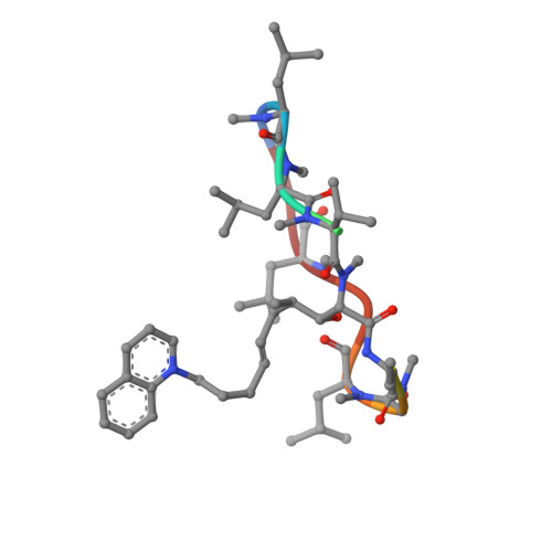
Crystal structure of cyclophilin D in complex with CsA analogue, JW47.


Experimental Data Snapshot

  • Method: X-RAY DIFFRACTION
  • Resolution: 1.25 Å
  • R-Value Free: 0.131 
  • R-Value Work: 0.098 
  • R-Value Observed: 0.100 

Starting Model: experimental
View more details

wwPDB Validation   3D Report Full Report


This is version 1.3 of the entry. See complete history


Literature

Selective Inhibition of the Mitochondrial Permeability Transition Pore Protects Against Neuro-Degeneration in Experimental Multiple Sclerosis.

Warne, J.Pryce, G.Hill, J.Shi, X.Lenneras, F.Puentes, F.Kip, M.Hilditch, L.Walker, P.Simone, M.I.Chan, A.W.E.Towers, G.J.Coker, A.R.Duchen, M.R.Szabadkai, G.Baker, D.Selwood, D.L.

(2016) J Biol Chem 291: 4356

  • DOI: https://doi.org/10.1074/jbc.M115.700385
  • Primary Citation of Related Structures:  
    5A0E

  • PubMed Abstract: 

    The mitochondrial permeability transition pore is a recognized drug target for neurodegenerative conditions such as multiple sclerosis and for ischemia-reperfusion injury in the brain and heart. The peptidylprolyl isomerase, cyclophilin D (CypD, PPIF), is a positive regulator of the pore, and genetic down-regulation or knock-out improves outcomes in disease models. Current inhibitors of peptidylprolyl isomerases show no selectivity between the tightly conserved cyclophilin paralogs and exhibit significant off-target effects, immunosuppression, and toxicity. We therefore designed and synthesized a new mitochondrially targeted CypD inhibitor, JW47, using a quinolinium cation tethered to cyclosporine. X-ray analysis was used to validate the design concept, and biological evaluation revealed selective cellular inhibition of CypD and the permeability transition pore with reduced cellular toxicity compared with cyclosporine. In an experimental autoimmune encephalomyelitis disease model of neurodegeneration in multiple sclerosis, JW47 demonstrated significant protection of axons and improved motor assessments with minimal immunosuppression. These findings suggest that selective CypD inhibition may represent a viable therapeutic strategy for MS and identify quinolinium as a mitochondrial targeting group for in vivo use.


  • Organizational Affiliation

    From the Wolfson Institute for Biomedical Research, University College London, Gower Street, London WC1E 6BT, United Kingdom.


Macromolecules
Find similar proteins by:  (by identity cutoff)  |  3D Structure
Entity ID: 1
MoleculeChains Sequence LengthOrganismDetailsImage
PEPTIDYL-PROLYL CIS-TRANS ISOMERASE F, MITOCHONDRIAL
A, B
165Homo sapiensMutation(s): 1 
EC: 5.2.1.8
UniProt & NIH Common Fund Data Resources
Find proteins for P30405 (Homo sapiens)
Explore P30405 
Go to UniProtKB:  P30405
PHAROS:  P30405
GTEx:  ENSG00000108179 
Entity Groups  
Sequence Clusters30% Identity50% Identity70% Identity90% Identity95% Identity100% Identity
UniProt GroupP30405
Sequence Annotations
Expand
  • Reference Sequence

Find similar proteins by:  Sequence   |   3D Structure  

Entity ID: 2
MoleculeChains Sequence LengthOrganismDetailsImage
JW47C,
D [auth E]
11Cylindrocarpon lucidumMutation(s): 0 
Entity Groups  
Sequence Clusters30% Identity50% Identity70% Identity90% Identity95% Identity100% Identity
Sequence Annotations
Expand
  • Reference Sequence
Small Molecules
Modified Residues  4 Unique
IDChains TypeFormula2D DiagramParent
ABA
Query on ABA
C,
D [auth E]
L-PEPTIDE LINKINGC4 H9 N O2ALA
MLE
Query on MLE
C,
D [auth E]
L-PEPTIDE LINKINGC7 H15 N O2LEU
MVA
Query on MVA
C,
D [auth E]
L-PEPTIDE LINKINGC6 H13 N O2VAL
SAR
Query on SAR
C,
D [auth E]
PEPTIDE LINKINGC3 H7 N O2GLY
Experimental Data & Validation

Experimental Data

  • Method: X-RAY DIFFRACTION
  • Resolution: 1.25 Å
  • R-Value Free: 0.131 
  • R-Value Work: 0.098 
  • R-Value Observed: 0.100 
  • Space Group: P 21 21 21
Unit Cell:
Length ( Å )Angle ( ˚ )
a = 38.12α = 90
b = 69.51β = 90
c = 109.17γ = 90
Software Package:
Software NamePurpose
REFMACrefinement
XDSdata reduction
XSCALEdata scaling
PHASERphasing

Structure Validation

View Full Validation Report


Entry History 

Revision History  (Full details and data files)

  • Version 1.0: 2015-12-30
    Type: Initial release
  • Version 1.1: 2016-03-16
    Changes: Database references
  • Version 1.2: 2019-05-08
    Changes: Data collection, Derived calculations, Experimental preparation, Other
  • Version 1.3: 2024-01-10
    Changes: Data collection, Database references, Derived calculations, Other, Refinement description