4WW0

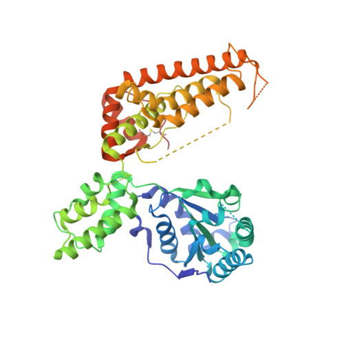
Truncated FtsH from A. aeolicus


Experimental Data Snapshot

  • Method: X-RAY DIFFRACTION
  • Resolution: 2.96 Å
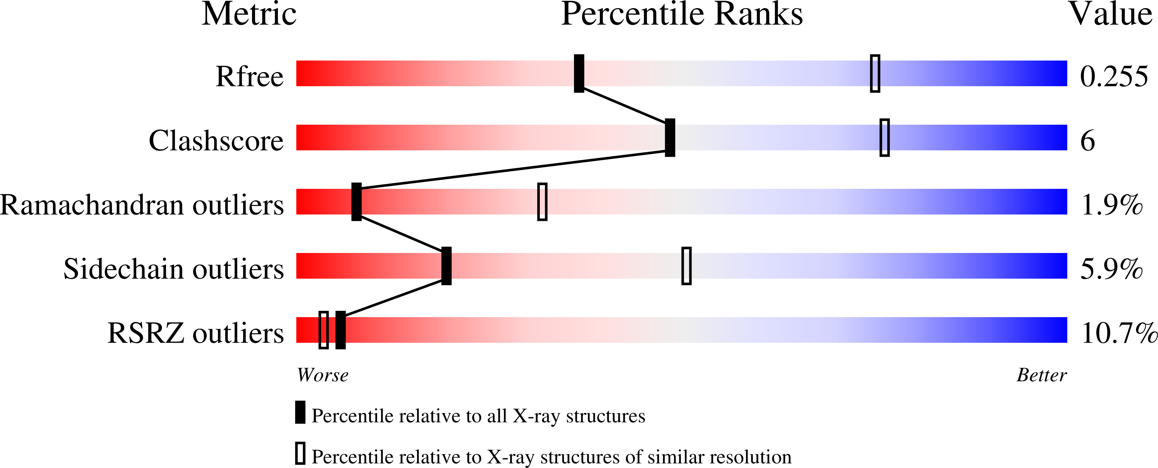
  • R-Value Free: 0.248 
  • R-Value Work: 0.209 
  • R-Value Observed: 0.211 

Starting Models: experimental
View more details

wwPDB Validation   3D Report Full Report


Ligand Structure Quality Assessment 


This is version 1.3 of the entry. See complete history


Literature

The structure of Aquifex aeolicus FtsH in the ADP-bound state reveals a C2-symmetric hexamer.

Vostrukhina, M.Popov, A.Brunstein, E.Lanz, M.A.Baumgartner, R.Bieniossek, C.Schacherl, M.Baumann, U.

(2015) Acta Crystallogr D Biol Crystallogr 71: 1307-1318

  • DOI: https://doi.org/10.1107/S1399004715005945
  • Primary Citation of Related Structures:  
    4WW0, 4Z8X

  • PubMed Abstract: 

    The crystal structure of a truncated, soluble quadruple mutant of FtsH from Aquifex aeolicus comprising the AAA and protease domains has been determined at 2.96 Å resolution in space group I222. The protein crystallizes as a hexamer, with the protease domain forming layers in the ab plane. Contacts between these layers are mediated by the AAA domains. These are highly disordered in one crystal form, but are clearly visible in a related form with a shorter c axis. Here, adenosine diphosphate (ADP) is bound to each subunit and the AAA ring exhibits twofold symmetry. The arrangement is different from the ADP-bound state of an analogously truncated, soluble FtsH construct from Thermotoga maritima. The pore is completely closed and the phenylalanine residues in the pore line a contiguous path. The protease hexamer is very similar to those described for other FtsH structures. To resolve certain open issues regarding a conserved glycine in the linker between the AAA and protease domains, as well as the active-site switch β-strand, mutations have been introduced in the full-length membrane-bound protein. Activity analysis of these point mutants reveals the crucial importance of these residues for proteolytic activity and is in accord with previous interpretation of the active-site switch and the importance of the linker glycine residue.


  • Organizational Affiliation

    Department of Chemistry and Biochemistry, University of Bern, Freiestrasse 3, Bern, CH-3012, Switzerland.


Macromolecules
Find similar proteins by:  (by identity cutoff)  |  3D Structure
Entity ID: 1
MoleculeChains Sequence LengthOrganismDetailsImage
ATP-dependent zinc metalloprotease FtsH
A, B, C
497Aquifex aeolicus VF5Mutation(s): 5 
Gene Names: ftsHaq_936
EC: 3.4.24
Membrane Entity: Yes 
UniProt
Find proteins for O67077 (Aquifex aeolicus (strain VF5))
Explore O67077 
Go to UniProtKB:  O67077
Entity Groups  
Sequence Clusters30% Identity50% Identity70% Identity90% Identity95% Identity100% Identity
UniProt GroupO67077
Sequence Annotations
Expand
  • Reference Sequence
Experimental Data & Validation

Experimental Data

  • Method: X-RAY DIFFRACTION
  • Resolution: 2.96 Å
  • R-Value Free: 0.248 
  • R-Value Work: 0.209 
  • R-Value Observed: 0.211 
  • Space Group: I 2 2 2
Unit Cell:
Length ( Å )Angle ( ˚ )
a = 138.23α = 90
b = 162.26β = 90
c = 170.17γ = 90
Software Package:
Software NamePurpose
BUSTERrefinement
XDSdata reduction
XSCALEdata scaling
PHASERphasing

Structure Validation

View Full Validation Report


Ligand Structure Quality Assessment 


Entry History & Funding Information

Deposition Data


Funding OrganizationLocationGrant Number
Swiss National Science FoundationSwitzerland31003A-120174
Swiss National Science FoundationSwitzerland3100A0-108262

Revision History  (Full details and data files)

  • Version 1.0: 2015-05-06
    Type: Initial release
  • Version 1.1: 2015-06-03
    Changes: Database references
  • Version 1.2: 2015-06-17
    Changes: Database references
  • Version 1.3: 2024-01-10
    Changes: Data collection, Database references, Derived calculations, Refinement description