Autoinhibitory mechanism and activity-related structural changes in a mycobacterial adenylyl cyclase
Barathy, D.V., Bharambe, N.G., Syed, W., Zaveri, A., Visweswariah, S.S., Cola sigmaf o, M., Misquith, S., Suguna, K.(2015) J Struct Biol 190: 304-313
- PubMed: 25916753
- DOI: https://doi.org/10.1016/j.jsb.2015.04.013
- Primary Citation of Related Structures:
4WP3, 4WP8, 4WP9, 4WPA - PubMed Abstract:
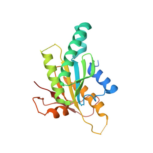
An adenylyl cyclase from Mycobacterium avium, Ma1120, is a functional orthologue of a pseudogene Rv1120c from Mycobacterium tuberculosis. We report the crystal structure of Ma1120 in a monomeric form and its truncated construct as a dimer. Ma1120 exists as a monomer in solution and crystallized as a monomer in the absence of substrate or inhibitor. An additional α-helix present at the N-terminus of the monomeric structure blocks the active site by interacting with the substrate binding residues and occupying the dimer interface region. However, the enzyme has been found to be active in solution, indicating the movement of the helix away from the interface to facilitate the formation of active dimers in conditions favourable for catalysis. Thus, the N-terminal helix of Ma1120 keeps the enzyme in an autoinhibited state when it is not active. Deletion of this helix enabled us to crystallize the molecule as an active homodimer in the presence of a P-site inhibitor 2',5'-dideoxy-3'-ATP, or pyrophosphate along with metal ions. The substrate specifying lysine residue plays a dual role of interacting with the substrate and stabilizing the dimer. The dimerization loop region harbouring the second substrate specifying residue, an aspartate, shows significant differences in conformation and position between the monomeric and dimeric structures. Thus, this study has not only revealed that significant structural transitions are required for the interconversion of the inactive and the active forms of the enzyme, but also provided precise nature of these transitions.
Organizational Affiliation:
Molecular Biophysics Unit, Indian Institute of Science, Bangalore 560 012, India.


