4WND

Crystal structure of the TPR domain of LGN in complex with Frmpd4/Preso1 at 1.5 Angstrom resolution


Experimental Data Snapshot

  • Method: X-RAY DIFFRACTION
  • Resolution: 1.50 Å
  • R-Value Free: 0.182 
  • R-Value Work: 0.163 
  • R-Value Observed: 0.164 

Starting Model: experimental
View more details

wwPDB Validation   3D Report Full Report


This is version 1.2 of the entry. See complete history


Literature

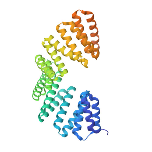
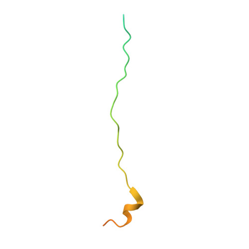
Structural basis for the recognition of the scaffold protein Frmpd4/Preso1 by the TPR domain of the adaptor protein LGN

Takayanagi, H.Yuzawa, S.Sumimoto, H.

(2015) Acta Crystallogr F Struct Biol Commun 71: 175-183

  • DOI: https://doi.org/10.1107/S2053230X14028143
  • Primary Citation of Related Structures:  
    4WND, 4WNE, 4WNF, 4WNG

  • PubMed Abstract: 

    The adaptor protein LGN interacts via the N-terminal domain comprising eight tetratricopeptide-repeat (TPR) motifs with its partner proteins mInsc, NuMA, Frmpd1 and Frmpd4 in a mutually exclusive manner. Here, the crystal structure of the LGN TPR domain in complex with human Frmpd4 is described at 1.5 Å resolution. In the complex, the LGN-binding region of Frmpd4 (amino-acid residues 990-1011) adopts an extended structure that runs antiparallel to LGN along the concave surface of the superhelix formed by the TPR motifs. Comparison with the previously determined structures of the LGN-Frmpd1, LGN-mInsc and LGN-NuMA complexes reveals that these partner proteins interact with LGN TPR1-6 via a common core binding region with consensus sequence (E/Q)XEX4-5(E/D/Q)X1-2(K/R)X0-1(V/I). In contrast to Frmpd1, Frmpd4 makes additional contacts with LGN via regions N- and C-terminal to the core sequence. The N-terminal extension is replaced by a specific α-helix in mInsc, which drastically increases the direct contacts with LGN TPR7/8, consistent with the higher affinity of mInsc for LGN. A crystal structure of Frmpd4-bound LGN in an oxidized form is also reported, although oxidation does not appear to strongly affect the interaction with Frmpd4.


  • Organizational Affiliation

    Department of Biochemistry, Kyushu University Graduate School of Medical Sciences, 3-1-1 Maidashi, Higashi-ku, Fukuoka 812-8582, Japan.


Macromolecules
Find similar proteins by:  (by identity cutoff)  |  3D Structure
Entity ID: 1
MoleculeChains Sequence LengthOrganismDetailsImage
G-protein-signaling modulator 2406Homo sapiensMutation(s): 0 
Gene Names: GPSM2LGN
UniProt & NIH Common Fund Data Resources
Find proteins for P81274 (Homo sapiens)
Explore P81274 
Go to UniProtKB:  P81274
GTEx:  ENSG00000121957 
Entity Groups  
Sequence Clusters30% Identity50% Identity70% Identity90% Identity95% Identity100% Identity
UniProt GroupP81274
Sequence Annotations
Expand
  • Reference Sequence
Find similar proteins by:  (by identity cutoff)  |  3D Structure
Entity ID: 2
MoleculeChains Sequence LengthOrganismDetailsImage
FERM and PDZ domain-containing protein 453Homo sapiensMutation(s): 0 
Gene Names: FRMPD4KIAA0316PDZD10PDZK10
UniProt & NIH Common Fund Data Resources
Find proteins for Q14CM0 (Homo sapiens)
Explore Q14CM0 
Go to UniProtKB:  Q14CM0
PHAROS:  Q14CM0
GTEx:  ENSG00000169933 
Entity Groups  
Sequence Clusters30% Identity50% Identity70% Identity90% Identity95% Identity100% Identity
UniProt GroupQ14CM0
Sequence Annotations
Expand
  • Reference Sequence
Small Molecules
Ligands 4 Unique
IDChains Name / Formula / InChI Key2D Diagram3D Interactions
PGE
Query on PGE

Download Ideal Coordinates CCD File 
O [auth A]TRIETHYLENE GLYCOL
C6 H14 O4
ZIBGPFATKBEMQZ-UHFFFAOYSA-N
PEG
Query on PEG

Download Ideal Coordinates CCD File 
J [auth A],
K [auth A],
L [auth A],
M [auth A],
N [auth A]
DI(HYDROXYETHYL)ETHER
C4 H10 O3
MTHSVFCYNBDYFN-UHFFFAOYSA-N
EDO
Query on EDO

Download Ideal Coordinates CCD File 
P [auth A]
Q [auth A]
R [auth A]
S [auth A]
T [auth A]
P [auth A],
Q [auth A],
R [auth A],
S [auth A],
T [auth A],
U [auth A]
1,2-ETHANEDIOL
C2 H6 O2
LYCAIKOWRPUZTN-UHFFFAOYSA-N
SCN
Query on SCN

Download Ideal Coordinates CCD File 
C [auth A]
D [auth A]
E [auth A]
F [auth A]
G [auth A]
C [auth A],
D [auth A],
E [auth A],
F [auth A],
G [auth A],
H [auth A],
I [auth A]
THIOCYANATE ION
C N S
ZMZDMBWJUHKJPS-UHFFFAOYSA-M
Experimental Data & Validation

Experimental Data

  • Method: X-RAY DIFFRACTION
  • Resolution: 1.50 Å
  • R-Value Free: 0.182 
  • R-Value Work: 0.163 
  • R-Value Observed: 0.164 
  • Space Group: P 1 21 1
Unit Cell:
Length ( Å )Angle ( ˚ )
a = 45.487α = 90
b = 64.786β = 102.33
c = 69.118γ = 90
Software Package:
Software NamePurpose
HKL-2000data reduction
HKL-2000data scaling
MOLREPphasing
Cootmodel building
REFMACrefinement
PDB_EXTRACTdata extraction

Structure Validation

View Full Validation Report


Entry History 

Deposition Data

Revision History  (Full details and data files)

  • Version 1.0: 2015-09-16
    Type: Initial release
  • Version 1.1: 2020-02-05
    Changes: Data collection, Derived calculations, Refinement description
  • Version 1.2: 2023-11-08
    Changes: Data collection, Database references, Refinement description