4V27

Structure of the GH99 endo-alpha-mannanase from Bacteroides xylanisolvens in complex with mannose-alpha-1,3-isofagomine


Experimental Data Snapshot

  • Method: X-RAY DIFFRACTION
  • Resolution: 1.90 Å
  • R-Value Free: 0.188 
  • R-Value Work: 0.151 
  • R-Value Observed: 0.152 

Starting Model: experimental
View more details

wwPDB Validation   3D Report Full Report


Ligand Structure Quality Assessment 


This is version 1.4 of the entry. See complete history


Literature

Structural and Kinetic Dissection of the Endo-Alpha-1,2-Mannanase Activity of Bacterial Gh99 Glycoside Hydrolases from Bacteroides Spp.

Hakki, Z.Thompson, A.J.Bellmaine, S.Speciale, G.Davies, G.J.Williams, S.J.

(2015) Chemistry 21: 1966

  • DOI: https://doi.org/10.1002/chem.201405539
  • Primary Citation of Related Structures:  
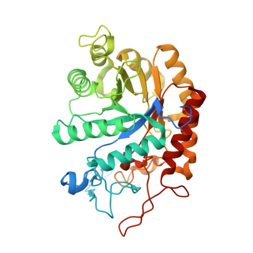
    4V27, 4V28

  • PubMed Abstract: 

    Glycoside hydrolase family 99 (GH99) was created to categorize sequence-related glycosidases possessing endo-α-mannosidase activity: the cleavage of mannosidic linkages within eukaryotic N-glycan precursors (Glc1-3 Man9 GlcNAc2 ), releasing mono-, di- and triglucosylated-mannose (Glc1-3 -1,3-Man). GH99 family members have recently been implicated in the ability of Bacteroides spp., present within the gut microbiota, to metabolize fungal cell wall α-mannans, releasing α-1,3-mannobiose by hydrolysing αMan-1,3-αMan→1,2-αMan-1,2-αMan sequences within branches off the main α-1,6-mannan backbone. We report the development of a series of substrates and inhibitors, which we use to kinetically and structurally characterise this novel endo-α-1,2-mannanase activity of bacterial GH99 enzymes from Bacteroides thetaiotaomicron and xylanisolvens. These data reveal an approximate 5 kJ mol(-1) preference for mannose-configured substrates in the -2 subsite (relative to glucose), which inspired the development of a new inhibitor, α-mannopyranosyl-1,3-isofagomine (ManIFG), the most potent (bacterial) GH99 inhibitor reported to date. X-ray structures of ManIFG or a substrate in complex with wild-type or inactive mutants, respectively, of B. xylanisolvens GH99 reveal the structural basis for binding to D-mannose- rather than D-glucose-configured substrates.


  • Organizational Affiliation

    School of Chemistry and Bio21 Molecular Science and Biotechnology Institute, University of Melbourne, Parkville, Victoria 3010 (Australia).


Macromolecules
Find similar proteins by:  (by identity cutoff)  |  3D Structure
Entity ID: 1
MoleculeChains Sequence LengthOrganismDetailsImage
GLYCOSYL HYDROLASE FAMILY 71380Bacteroides xylanisolvens XB1AMutation(s): 0 
EC: 3.2.1.130
UniProt
Find proteins for D6D1V7 (Bacteroides xylanisolvens XB1A)
Explore D6D1V7 
Go to UniProtKB:  D6D1V7
Entity Groups  
Sequence Clusters30% Identity50% Identity70% Identity90% Identity95% Identity100% Identity
UniProt GroupD6D1V7
Sequence Annotations
Expand
  • Reference Sequence
Experimental Data & Validation

Experimental Data

  • Method: X-RAY DIFFRACTION
  • Resolution: 1.90 Å
  • R-Value Free: 0.188 
  • R-Value Work: 0.151 
  • R-Value Observed: 0.152 
  • Space Group: I 4
Unit Cell:
Length ( Å )Angle ( ˚ )
a = 108.28α = 90
b = 108.28β = 90
c = 67.62γ = 90
Software Package:
Software NamePurpose
REFMACrefinement
MOSFLMdata reduction
SCALAdata scaling
REFMACphasing

Structure Validation

View Full Validation Report


Ligand Structure Quality Assessment 


Entry History 

Deposition Data

Revision History  (Full details and data files)

  • Version 1.0: 2014-12-24
    Type: Initial release
  • Version 1.1: 2015-02-04
    Changes: Database references
  • Version 1.2: 2015-07-15
    Changes: Non-polymer description
  • Version 1.3: 2020-07-29
    Type: Remediation
    Reason: Carbohydrate remediation
    Changes: Data collection, Derived calculations, Other, Structure summary
  • Version 1.4: 2024-01-10
    Changes: Data collection, Database references, Refinement description, Structure summary