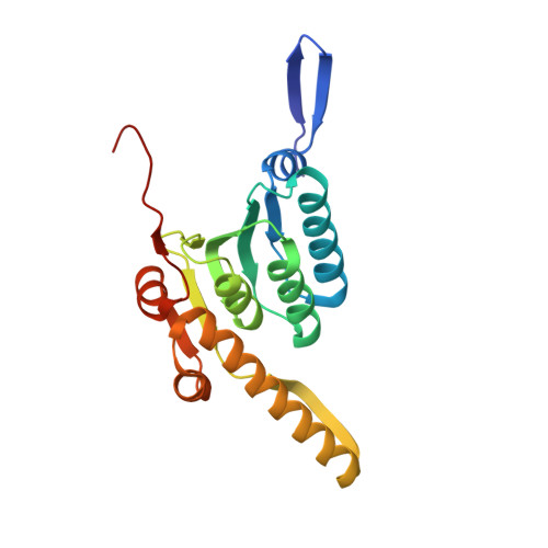
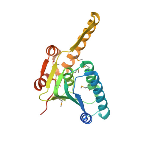
4U0G

Crystal Structure of M. tuberculosis ClpP1P2 bound to ADEP and agonist


Experimental Data Snapshot

  • Method: X-RAY DIFFRACTION
  • Resolution: 3.20 Å
  • R-Value Free: 0.222 
  • R-Value Work: 0.190 
  • R-Value Observed: 0.192 

wwPDB Validation   3D Report Full Report


Ligand Structure Quality Assessment 


This is version 2.0 of the entry. See complete history


Literature

Crystal structure of Mycobacterium tuberculosis ClpP1P2 suggests a model for peptidase activation by AAA+ partner binding and substrate delivery.

Schmitz, K.R.Carney, D.W.Sello, J.K.Sauer, R.T.

(2014) Proc Natl Acad Sci U S A 111: E4587-E4595

  • DOI: https://doi.org/10.1073/pnas.1417120111
  • Primary Citation of Related Structures:  
    4U0G, 4U0H

  • PubMed Abstract: 

    Caseinolytic peptidase P (ClpP), a double-ring peptidase with 14 subunits, collaborates with ATPases associated with diverse activities (AAA+) partners to execute ATP-dependent protein degradation. Although many ClpP enzymes self-assemble into catalytically active homo-tetradecamers able to cleave small peptides, the Mycobacterium tuberculosis enzyme consists of discrete ClpP1 and ClpP2 heptamers that require a AAA+ partner and protein-substrate delivery or a peptide agonist to stabilize assembly of the active tetradecamer. Here, we show that cyclic acyldepsipeptides (ADEPs) and agonist peptides synergistically activate ClpP1P2 by mimicking AAA+ partners and substrates, respectively, and determine the structure of the activated complex. Our studies establish the basis of heteromeric ClpP1P2 assembly and function, reveal tight coupling between the conformations of each ring, show that ADEPs bind only to one ring but appear to open the axial pores of both rings, provide a foundation for rational drug development, and suggest strategies for studying the roles of individual ClpP1 and ClpP2 rings in Clp-family proteolysis.


  • Organizational Affiliation

    Department of Biology, Massachusetts Institute of Technology, Cambridge, MA 02139; and.


Macromolecules
Find similar proteins by:  (by identity cutoff)  |  3D Structure
Entity ID: 1
MoleculeChains Sequence LengthOrganismDetailsImage
ATP-dependent Clp protease proteolytic subunit 2
A, B, C, D, E
A, B, C, D, E, F, G, O, P, Q, R, S, T, U
202Mycobacterium tuberculosis H37RvMutation(s): 0 
Gene Names: clpP2Rv2460cMTV008.16c
EC: 3.4.21.92
UniProt
Find proteins for P9WPC3 (Mycobacterium tuberculosis (strain ATCC 25618 / H37Rv))
Explore P9WPC3 
Go to UniProtKB:  P9WPC3
Entity Groups  
Sequence Clusters30% Identity50% Identity70% Identity90% Identity95% Identity100% Identity
UniProt GroupP9WPC3
Sequence Annotations
Expand
  • Reference Sequence
Find similar proteins by:  (by identity cutoff)  |  3D Structure
Entity ID: 2
MoleculeChains Sequence LengthOrganismDetailsImage
ATP-dependent Clp protease proteolytic subunit 1194Mycobacterium tuberculosis H37RvMutation(s): 0 
Gene Names: clpP1clpPRv2461cMTV008.17c
EC: 3.4.21.92
UniProt
Find proteins for P9WPC5 (Mycobacterium tuberculosis (strain ATCC 25618 / H37Rv))
Explore P9WPC5 
Go to UniProtKB:  P9WPC5
Entity Groups  
Sequence Clusters30% Identity50% Identity70% Identity90% Identity95% Identity100% Identity
UniProt GroupP9WPC5
Sequence Annotations
Expand
  • Reference Sequence

Find similar proteins by:  Sequence   |   3D Structure  

Entity ID: 3
MoleculeChains Sequence LengthOrganismDetailsImage
ADEP-2B5Me7synthetic constructMutation(s): 0 
Sequence Annotations
Expand
  • Reference Sequence
Small Molecules
Ligands 3 Unique
IDChains Name / Formula / InChI Key2D Diagram3D Interactions
ZIL
Query on ZIL

Download Ideal Coordinates CCD File 
AC [auth M]
BD [auth T]
CC [auth N]
DB [auth D]
DC [auth O]
AC [auth M],
BD [auth T],
CC [auth N],
DB [auth D],
DC [auth O],
ED [auth U],
GB [auth E],
HC [auth P],
HD [auth V],
ID [auth W],
KD [auth X],
LD [auth Y],
MB [auth F],
MC [auth Q],
MD [auth Z],
ND [auth a],
OD [auth b],
PB [auth G],
QA [auth A],
QC [auth R],
TA [auth B],
TB [auth H],
UB [auth I],
VC [auth S],
WB [auth J],
XA [auth C],
YB [auth K],
ZB [auth L]
N-[(benzyloxy)carbonyl]-L-isoleucyl-L-leucine
C20 H30 N2 O5
BSRAGXJNZJMFMY-XIRDDKMYSA-N
SO4
Query on SO4

Download Ideal Coordinates CCD File 
AB [auth C]
BB [auth C]
BC [auth M]
CD [auth T]
EB [auth D]
AB [auth C],
BB [auth C],
BC [auth M],
CD [auth T],
EB [auth D],
EC [auth O],
FC [auth O],
FD [auth U],
HB [auth E],
IB [auth E],
IC [auth P],
JB [auth E],
JC [auth P],
JD [auth W],
KB [auth E],
KC [auth P],
NB [auth F],
NC [auth Q],
OC [auth Q],
PD [auth p],
QB [auth G],
RA [auth A],
RB [auth G],
RC [auth R],
SC [auth R],
TC [auth R],
UA [auth B],
VB [auth I],
WC [auth S],
XB [auth J],
XC [auth S],
YA [auth C],
YC [auth S],
ZA [auth C],
ZC [auth S]
SULFATE ION
O4 S
QAOWNCQODCNURD-UHFFFAOYSA-L
DMS
Query on DMS

Download Ideal Coordinates CCD File 
AD [auth S]
CB [auth C]
DD [auth T]
FB [auth D]
GC [auth O]
AD [auth S],
CB [auth C],
DD [auth T],
FB [auth D],
GC [auth O],
GD [auth U],
LB [auth E],
LC [auth P],
OB [auth F],
PC [auth Q],
SA [auth A],
SB [auth G],
UC [auth R],
VA [auth B],
WA [auth B]
DIMETHYL SULFOXIDE
C2 H6 O S
IAZDPXIOMUYVGZ-UHFFFAOYSA-N
Modified Residues  2 Unique
IDChains TypeFormula2D DiagramParent
MSE
Query on MSE
AA [auth a]
BA [auth b]
H
I
J
AA [auth a],
BA [auth b],
H,
I,
J,
K,
L,
M,
N,
V,
W,
X,
Y,
Z
L-PEPTIDE LINKINGC5 H11 N O2 SeMET
WFP
Query on WFP
CA [auth c]
DA [auth i]
EA [auth d]
FA [auth e]
GA [auth f]
CA [auth c],
DA [auth i],
EA [auth d],
FA [auth e],
GA [auth f],
HA [auth g],
IA [auth h],
JA [auth j],
KA [auth k],
LA [auth l],
MA [auth m],
NA [auth n],
OA [auth o],
PA [auth p]
L-PEPTIDE LINKINGC9 H9 F2 N O2PHE
Biologically Interesting Molecules (External Reference) 1 Unique
Entity ID: 3
IDChains NameType/Class2D Diagram3D Interactions
PRD_002111
Query on PRD_002111
CA [auth c]
DA [auth d]
EA [auth e]
FA [auth f]
GA [auth g]
CA [auth c],
DA [auth d],
EA [auth e],
FA [auth f],
GA [auth g],
HA [auth h],
IA [auth i],
JA [auth j],
KA [auth k],
LA [auth l],
MA [auth m],
NA [auth n],
OA [auth o],
PA [auth p]
ADEP-2B5MeCyclic depsipeptide / Antibiotic
Experimental Data & Validation

Experimental Data

  • Method: X-RAY DIFFRACTION
  • Resolution: 3.20 Å
  • R-Value Free: 0.222 
  • R-Value Work: 0.190 
  • R-Value Observed: 0.192 
  • Space Group: P 21 21 21
Unit Cell:
Length ( Å )Angle ( ˚ )
a = 154.748α = 90
b = 187.673β = 90
c = 294.029γ = 90
Software Package:
Software NamePurpose
PHENIXrefinement
HKL-2000data scaling
HKL-2000data reduction
Cootmodel building
PDB_EXTRACTdata extraction
PHASERphasing
DENZOdata reduction
SCALEPACKdata scaling

Structure Validation

View Full Validation Report


Ligand Structure Quality Assessment 


Entry History & Funding Information

Deposition Data


Funding OrganizationLocationGrant Number
National Institutes of Health/National Institute of General Medical Sciences (NIH/NIGMS)United StatesGM-101988

Revision History  (Full details and data files)

  • Version 1.0: 2014-10-08
    Type: Initial release
  • Version 1.1: 2014-10-15
    Changes: Database references
  • Version 1.2: 2014-11-12
    Changes: Database references
  • Version 1.3: 2017-09-06
    Changes: Author supporting evidence, Database references, Derived calculations, Other, Refinement description, Source and taxonomy
  • Version 1.4: 2019-12-25
    Changes: Author supporting evidence
  • Version 2.0: 2023-11-15
    Changes: Atomic model, Data collection, Database references, Derived calculations, Refinement description