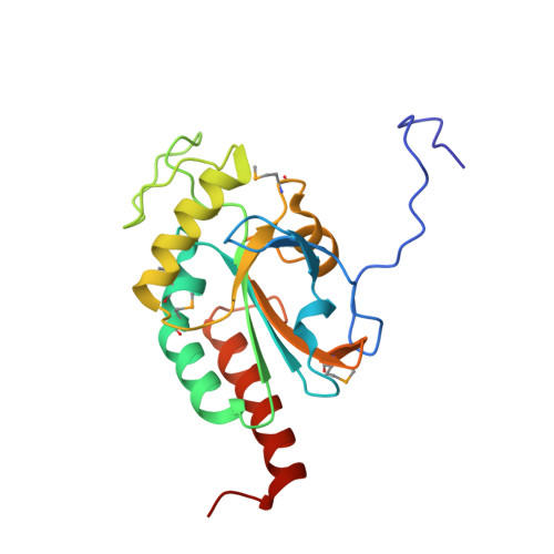
4TR3

Mouse iodothyronine deiodinase 3 catalytic core, SeMet-labeled active site mutant SeCys->Cys


Experimental Data Snapshot

  • Method: X-RAY DIFFRACTION
  • Resolution: 1.90 Å
  • R-Value Free: 0.227 
  • R-Value Work: 0.187 
  • R-Value Observed: 0.189 

wwPDB Validation   3D Report Full Report


This is version 1.2 of the entry. See complete history


Literature

Crystal structure of mammalian selenocysteine-dependent iodothyronine deiodinase suggests a peroxiredoxin-like catalytic mechanism.

Schweizer, U.Schlicker, C.Braun, D.Kohrle, J.Steegborn, C.

(2014) Proc Natl Acad Sci U S A 111: 10526

  • DOI: https://doi.org/10.1073/pnas.1323873111
  • Primary Citation of Related Structures:  
    4TR3, 4TR4

  • PubMed Abstract: 

    Local levels of active thyroid hormone (3,3',5-triiodothyronine) are controlled by the action of activating and inactivating iodothyronine deiodinase enzymes. Deiodinases are selenocysteine-dependent membrane proteins catalyzing the reductive elimination of iodide from iodothyronines through a poorly understood mechanism. We solved the crystal structure of the catalytic domain of mouse deiodinase 3 (Dio3), which reveals a close structural similarity to atypical 2-Cys peroxiredoxin(s) (Prx). The structure suggests a route for proton transfer to the substrate during deiodination and a Prx-related mechanism for subsequent recycling of the transiently oxidized enzyme. The proposed mechanism is supported by biochemical experiments and is consistent with the effects of mutations of conserved amino acids on Dio3 activity. Thioredoxin and glutaredoxin reduce the oxidized Dio3 at physiological concentrations, and dimerization appears to activate the enzyme by displacing an autoinhibitory loop from the iodothyronine binding site. Deiodinases apparently evolved from the ubiquitous Prx scaffold, and their structure and catalytic mechanism reconcile a plethora of partly conflicting data reported for these enzymes.


  • Organizational Affiliation

    Institut für Biochemie und Molekularbiologie, Rheinische Friedrich Wilhelms-Universität Bonn, 53115 Bonn, Germany; uschweiz@uni-bonn.de clemens.steegborn@uni-bayreuth.de.


Macromolecules
Find similar proteins by:  (by identity cutoff)  |  3D Structure
Entity ID: 1
MoleculeChains Sequence LengthOrganismDetailsImage
Type III iodothyronine deiodinase191Mus musculusMutation(s): 1 
Gene Names: Dio3
EC: 1.97.1.11 (PDB Primary Data), 1.21.99.3 (UniProt)
UniProt
Find proteins for Q91ZI8 (Mus musculus)
Explore Q91ZI8 
Go to UniProtKB:  Q91ZI8
Entity Groups  
Sequence Clusters30% Identity50% Identity70% Identity90% Identity95% Identity100% Identity
UniProt GroupQ91ZI8
Sequence Annotations
Expand
  • Reference Sequence
Small Molecules
Modified Residues  1 Unique
IDChains TypeFormula2D DiagramParent
MSE
Query on MSE
A
L-PEPTIDE LINKINGC5 H11 N O2 SeMET
Experimental Data & Validation

Experimental Data

  • Method: X-RAY DIFFRACTION
  • Resolution: 1.90 Å
  • R-Value Free: 0.227 
  • R-Value Work: 0.187 
  • R-Value Observed: 0.189 
  • Space Group: P 21 21 21
Unit Cell:
Length ( Å )Angle ( ˚ )
a = 48.92α = 90
b = 54.3β = 90
c = 66.61γ = 90
Software Package:
Software NamePurpose
REFMACrefinement
PDB_EXTRACTdata extraction
XDSdata reduction
SHELXphasing
ARPmodel building
Cootmodel building

Structure Validation

View Full Validation Report


Entry History 

Deposition Data

Revision History  (Full details and data files)

  • Version 1.0: 2014-07-23
    Type: Initial release
  • Version 1.1: 2015-02-04
    Changes: Data collection
  • Version 1.2: 2024-10-16
    Changes: Data collection, Database references, Structure summary