4QZD

Mouse Tdt, F405A mutant, in complex with a DSB substrate, C-C base pair


Experimental Data Snapshot

  • Method: X-RAY DIFFRACTION
  • Resolution: 2.70 Å
  • R-Value Free: 0.245 
  • R-Value Work: 0.200 
  • R-Value Observed: 0.202 

Starting Model: experimental
View more details

wwPDB Validation   3D Report Full Report


This is version 1.2 of the entry. See complete history


Literature

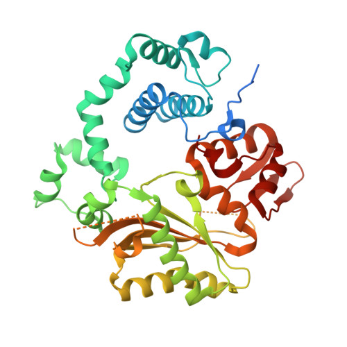
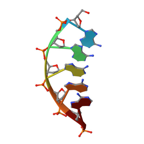
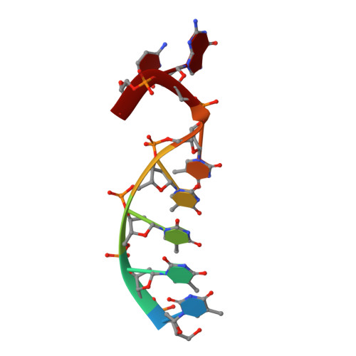
Structural basis for a novel mechanism of DNA bridging and alignment in eukaryotic DSB DNA repair.

Gouge, J.Rosario, S.Romain, F.Poitevin, F.Beguin, P.Delarue, M.

(2015) EMBO J 34: 1126-1142

  • DOI: https://doi.org/10.15252/embj.201489643
  • Primary Citation of Related Structures:  
    4QZ8, 4QZ9, 4QZA, 4QZB, 4QZC, 4QZD, 4QZE, 4QZF, 4QZG, 4QZH, 4QZI

  • PubMed Abstract: 

    Eukaryotic DNA polymerase mu of the PolX family can promote the association of the two 3'-protruding ends of a DNA double-strand break (DSB) being repaired (DNA synapsis) even in the absence of the core non-homologous end-joining (NHEJ) machinery. Here, we show that terminal deoxynucleotidyltransferase (TdT), a closely related PolX involved in V(D)J recombination, has the same property. We solved its crystal structure with an annealed DNA synapsis containing one micro-homology (MH) base pair and one nascent base pair. This structure reveals how the N-terminal domain and Loop 1 of Tdt cooperate for bridging the two DNA ends, providing a templating base in trans and limiting the MH search region to only two base pairs. A network of ordered water molecules is proposed to assist the incorporation of any nucleotide independently of the in trans templating base. These data are consistent with a recent model that explains the statistics of sequences synthesized in vivo by Tdt based solely on this dinucleotide step. Site-directed mutagenesis and functional tests suggest that this structural model is also valid for Pol mu during NHEJ.


  • Organizational Affiliation

    Unité de Dynamique Structurale des Macromolécules, Institut Pasteur, UMR 3528 du C.N.R.S., Paris, France.


Macromolecules

Find similar proteins by:  (by identity cutoff)  |  3D Structure
Entity ID: 1
MoleculeChains Sequence LengthOrganismDetailsImage
DNA nucleotidylexotransferase400Mus musculusMutation(s): 1 
Gene Names: DnttTdt
EC: 2.7.7.31
UniProt & NIH Common Fund Data Resources
Find proteins for P09838 (Mus musculus)
Explore P09838 
Go to UniProtKB:  P09838
IMPC:  MGI:98659
Entity Groups  
Sequence Clusters30% Identity50% Identity70% Identity90% Identity95% Identity100% Identity
UniProt GroupP09838
Sequence Annotations
Expand
  • Reference Sequence

Find similar nucleic acids by:  Sequence   |   3D Structure  

Entity ID: 2
MoleculeChains LengthOrganismImage
5'-D(*AP*AP*AP*AP*AP*C)-3'B [auth D],
D [auth U]
6N/A
Sequence Annotations
Expand
  • Reference Sequence

Find similar nucleic acids by:  Sequence   |   3D Structure  

Entity ID: 3
MoleculeChains LengthOrganismImage
5'-D(*TP*TP*TP*TP*TP*GP*C)-3'C [auth T]7N/A
Sequence Annotations
Expand
  • Reference Sequence
Experimental Data & Validation

Experimental Data

  • Method: X-RAY DIFFRACTION
  • Resolution: 2.70 Å
  • R-Value Free: 0.245 
  • R-Value Work: 0.200 
  • R-Value Observed: 0.202 
  • Space Group: C 1 2 1
Unit Cell:
Length ( Å )Angle ( ˚ )
a = 55.933α = 90
b = 74.92β = 98.43
c = 125.633γ = 90
Software Package:
Software NamePurpose
XDSdata scaling
PHASERphasing
BUSTERrefinement
XDSdata reduction
SCALAdata scaling

Structure Validation

View Full Validation Report


Entry History 

Deposition Data

Revision History  (Full details and data files)

  • Version 1.0: 2015-06-10
    Type: Initial release
  • Version 1.1: 2017-07-26
    Changes: Refinement description, Source and taxonomy
  • Version 1.2: 2023-09-20
    Changes: Data collection, Database references, Derived calculations, Refinement description