4QDC

Crystal structure of 3-ketosteroid-9-alpha-hydroxylase 5 (KshA5) from R. rhodochrous in complex with FE2/S2 (INORGANIC) CLUSTER


Experimental Data Snapshot

  • Method: X-RAY DIFFRACTION
  • Resolution: 1.90 Å
  • R-Value Free: 0.217 
  • R-Value Work: 0.187 
  • R-Value Observed: 0.188 

Starting Model: experimental
View more details

wwPDB Validation   3D Report Full Report


Ligand Structure Quality Assessment 


This is version 1.3 of the entry. See complete history


Literature

Substrate specificities and conformational flexibility of 3-ketosteroid 9 alpha-hydroxylases.

Penfield, J.S.Worrall, L.J.Strynadka, N.C.Eltis, L.D.

(2014) J Biol Chem 289: 25523-25536

  • DOI: https://doi.org/10.1074/jbc.M114.575886
  • Primary Citation of Related Structures:  
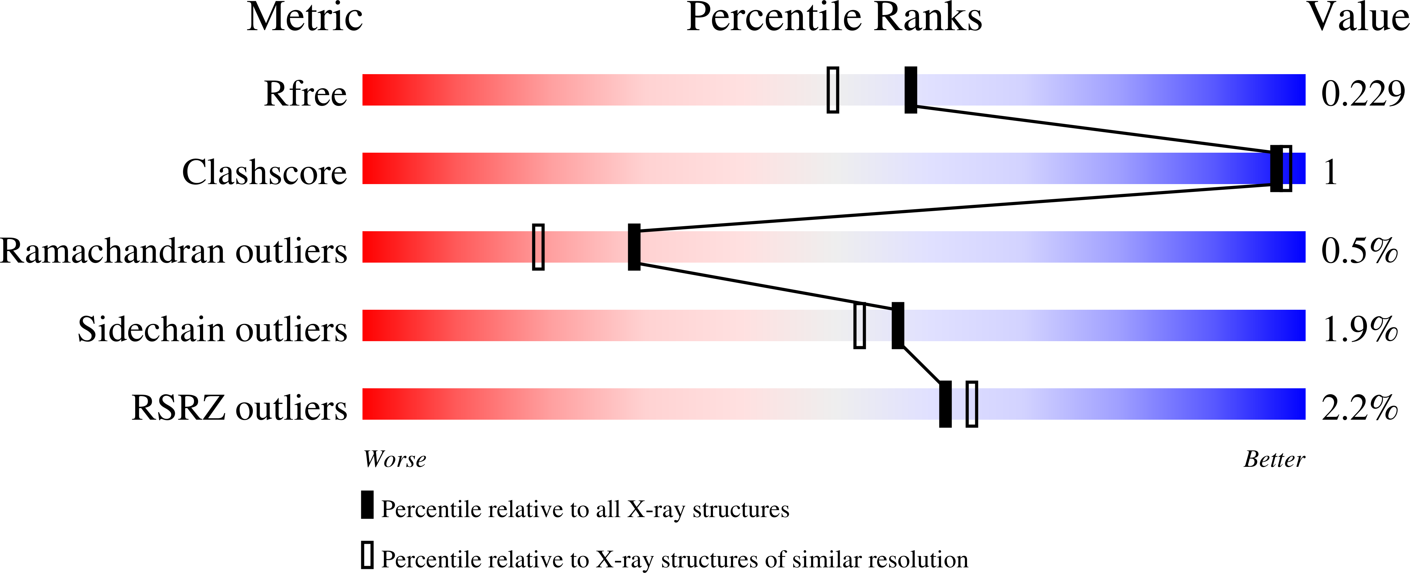
    4QCK, 4QDC, 4QDD, 4QDF

  • PubMed Abstract: 

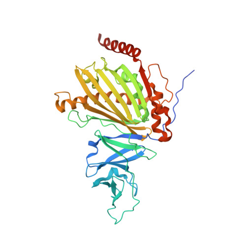
    KshA is the oxygenase component of 3-ketosteroid 9α-hydroxylase, a Rieske oxygenase involved in the bacterial degradation of steroids. Consistent with its role in bile acid catabolism, KshA1 from Rhodococcus rhodochrous DSM43269 had the highest apparent specificity (kcat/Km) for steroids with an isopropyl side chain at C17, such as 3-oxo-23,24-bisnorcholesta-1,4-diene-22-oate (1,4-BNC). By contrast, the KshA5 homolog had the highest apparent specificity for substrates with no C17 side chain (kcat/Km >10(5) s(-1) M(-1) for 4-estrendione, 5α-androstandione, and testosterone). Unexpectedly, substrates such as 4-androstene-3,17-dione (ADD) and 4-BNC displayed strong substrate inhibition (Ki S ∼100 μM). By comparison, the cholesterol-degrading KshAMtb from Mycobacterium tuberculosis had the highest specificity for CoA-thioesterified substrates. These specificities are consistent with differences in the catabolism of cholesterol and bile acids, respectively, in actinobacteria. X-ray crystallographic structures of the KshAMtb·ADD, KshA1·1,4-BNC-CoA, KshA5·ADD, and KshA5·1,4-BNC-CoA complexes revealed that the enzymes have very similar steroid-binding pockets with the substrate's C17 oriented toward the active site opening. Comparisons suggest Tyr-245 and Phe-297 are determinants of KshA1 specificity. All enzymes have a flexible 16-residue "mouth loop," which in some structures completely occluded the substrate-binding pocket from the bulk solvent. Remarkably, the catalytic iron and α-helices harboring its ligands were displaced up to 4.4 Å in the KshA5·substrate complexes as compared with substrate-free KshA, suggesting that Rieske oxygenases may have a dynamic nature similar to cytochrome P450.


  • Organizational Affiliation

    From the Departments of Biochemistry and Molecular Biology and.


Macromolecules
Find similar proteins by:  (by identity cutoff)  |  3D Structure
Entity ID: 1
MoleculeChains Sequence LengthOrganismDetailsImage
3-ketosteroid 9alpha-hydroxylase oxygenase390Rhodococcus rhodochrousMutation(s): 0 
Gene Names: kshakshA5
UniProt
Find proteins for F1CMY8 (Rhodococcus rhodochrous)
Explore F1CMY8 
Go to UniProtKB:  F1CMY8
Entity Groups  
Sequence Clusters30% Identity50% Identity70% Identity90% Identity95% Identity100% Identity
UniProt GroupF1CMY8
Sequence Annotations
Expand
  • Reference Sequence
Small Molecules
Ligands 6 Unique
IDChains Name / Formula / InChI Key2D Diagram3D Interactions
ASD
Query on ASD

Download Ideal Coordinates CCD File 
E [auth A]4-ANDROSTENE-3-17-DIONE
C19 H26 O2
AEMFNILZOJDQLW-QAGGRKNESA-N
PG4
Query on PG4

Download Ideal Coordinates CCD File 
G [auth A]TETRAETHYLENE GLYCOL
C8 H18 O5
UWHCKJMYHZGTIT-UHFFFAOYSA-N
FES
Query on FES

Download Ideal Coordinates CCD File 
B [auth A]FE2/S2 (INORGANIC) CLUSTER
Fe2 S2
NIXDOXVAJZFRNF-UHFFFAOYSA-N
SO4
Query on SO4

Download Ideal Coordinates CCD File 
D [auth A]SULFATE ION
O4 S
QAOWNCQODCNURD-UHFFFAOYSA-L
GOL
Query on GOL

Download Ideal Coordinates CCD File 
F [auth A]GLYCEROL
C3 H8 O3
PEDCQBHIVMGVHV-UHFFFAOYSA-N
FE
Query on FE

Download Ideal Coordinates CCD File 
C [auth A]FE (III) ION
Fe
VTLYFUHAOXGGBS-UHFFFAOYSA-N
Experimental Data & Validation

Experimental Data

  • Method: X-RAY DIFFRACTION
  • Resolution: 1.90 Å
  • R-Value Free: 0.217 
  • R-Value Work: 0.187 
  • R-Value Observed: 0.188 
  • Space Group: P 63
Unit Cell:
Length ( Å )Angle ( ˚ )
a = 162.183α = 90
b = 162.183β = 90
c = 46.969γ = 120
Software Package:
Software NamePurpose
Aimlessdata scaling
PHASERphasing
REFMACrefinement
PDB_EXTRACTdata extraction
MxDCdata collection
XDSdata reduction

Structure Validation

View Full Validation Report


Ligand Structure Quality Assessment 


Entry History 

Deposition Data

Revision History  (Full details and data files)

  • Version 1.0: 2014-07-30
    Type: Initial release
  • Version 1.1: 2014-10-01
    Changes: Database references
  • Version 1.2: 2017-11-22
    Changes: Refinement description
  • Version 1.3: 2023-09-20
    Changes: Data collection, Database references, Derived calculations, Refinement description