4Q5O

Crystal structure of EctD from S. alaskensis with 2-oxoglutarate and 5-hydroxyectoine


Experimental Data Snapshot

  • Method: X-RAY DIFFRACTION
  • Resolution: 2.64 Å
  • R-Value Free: 0.279 
  • R-Value Work: 0.206 
  • R-Value Observed: 0.210 

Starting Model: experimental
View more details

wwPDB Validation   3D Report Full Report


Ligand Structure Quality Assessment 


This is version 1.2 of the entry. See complete history


Literature

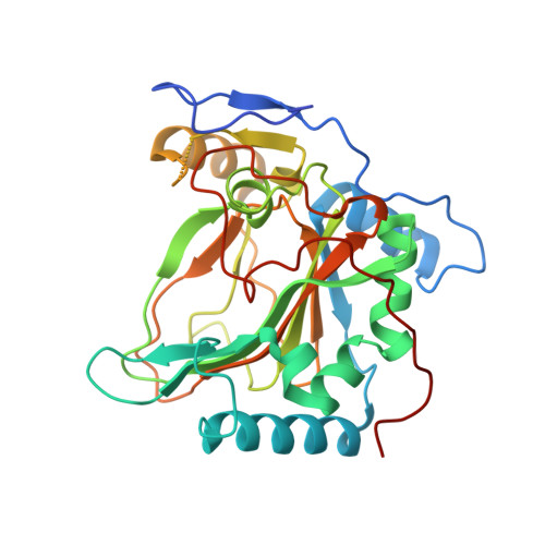
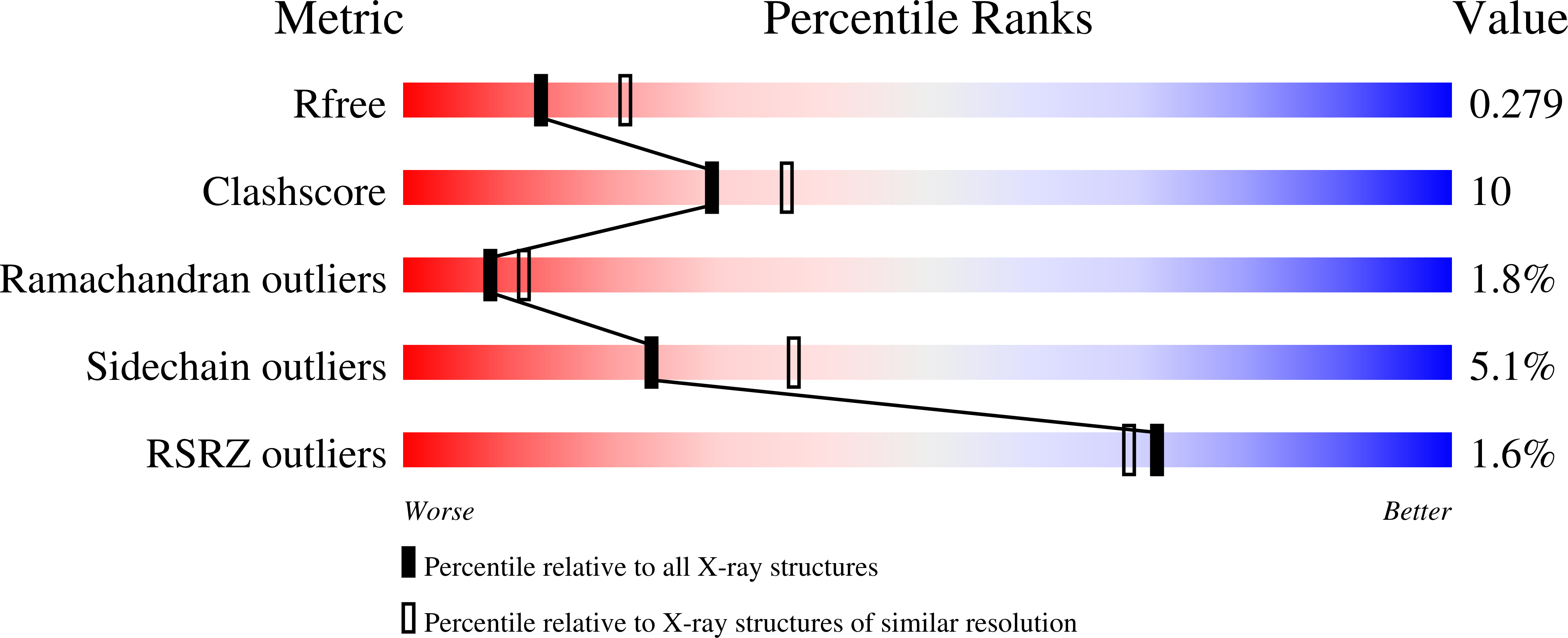
Crystal structure of the ectoine hydroxylase, a snapshot of the active site.

Hoppner, A.Widderich, N.Lenders, M.Bremer, E.Smits, S.H.

(2014) J Biol Chem 289: 29570-29583

  • DOI: https://doi.org/10.1074/jbc.M114.576769
  • Primary Citation of Related Structures:  
    4MHR, 4MHU, 4Q5O

  • PubMed Abstract: 

    Ectoine and its derivative 5-hydroxyectoine are compatible solutes that are widely synthesized by bacteria to cope physiologically with osmotic stress. They also serve as chemical chaperones and maintain the functionality of macromolecules. 5-Hydroxyectoine is produced from ectoine through a stereo-specific hydroxylation, an enzymatic reaction catalyzed by the ectoine hydroxylase (EctD). The EctD protein is a member of the non-heme-containing iron(II) and 2-oxoglutarate-dependent dioxygenase superfamily and is evolutionarily well conserved. We studied the ectoine hydroxylase from the cold-adapted marine ultra-microbacterium Sphingopyxis alaskensis (Sa) and found that the purified SaEctD protein is a homodimer in solution. We determined the SaEctD crystal structure in its apo-form, complexed with the iron catalyst, and in a form that contained iron, the co-substrate 2-oxoglutarate, and the reaction product of EctD, 5-hydroxyectoine. The iron and 2-oxoglutarate ligands are bound within the EctD active site in a fashion similar to that found in other members of the dioxygenase superfamily. 5-Hydroxyectoine, however, is coordinated by EctD in manner different from that found in high affinity solute receptor proteins operating in conjunction with microbial import systems for ectoines. Our crystallographic analysis provides a detailed view into the active site of the ectoine hydroxylase and exposes an intricate network of interactions between the enzyme and its ligands that collectively ensure the hydroxylation of the ectoine substrate in a position- and stereo-specific manner.


  • Organizational Affiliation

    From the X-ray Facility and Crystal Farm, Heinrich-Heine-University at Düsseldorf, D-40225 Düsseldorf.


Macromolecules
Find similar proteins by:  (by identity cutoff)  |  3D Structure
Entity ID: 1
MoleculeChains Sequence LengthOrganismDetailsImage
Ectoine hydroxylase
A, B
314Sphingopyxis alaskensis RB2256Mutation(s): 0 
Gene Names: Sala_2952
EC: 1.14.11.55
UniProt
Find proteins for Q1GNW5 (Sphingopyxis alaskensis (strain DSM 13593 / LMG 18877 / RB2256))
Explore Q1GNW5 
Go to UniProtKB:  Q1GNW5
Entity Groups  
Sequence Clusters30% Identity50% Identity70% Identity90% Identity95% Identity100% Identity
UniProt GroupQ1GNW5
Sequence Annotations
Expand
  • Reference Sequence
Small Molecules
Ligands 3 Unique
IDChains Name / Formula / InChI Key2D Diagram3D Interactions
6CS
Query on 6CS

Download Ideal Coordinates CCD File 
E [auth A],
H [auth B]
(4S,5S)-5-HYDROXY-2-METHYL-1,4,5,6-TETRAHYDROPYRIMIDINE-4-CARBOXYLIC ACID
C6 H10 N2 O3
KIIBBJKLKFTNQO-WHFBIAKZSA-N
AKG
Query on AKG

Download Ideal Coordinates CCD File 
D [auth A],
G [auth B]
2-OXOGLUTARIC ACID
C5 H6 O5
KPGXRSRHYNQIFN-UHFFFAOYSA-N
FE
Query on FE

Download Ideal Coordinates CCD File 
C [auth A],
F [auth B]
FE (III) ION
Fe
VTLYFUHAOXGGBS-UHFFFAOYSA-N
Experimental Data & Validation

Experimental Data

  • Method: X-RAY DIFFRACTION
  • Resolution: 2.64 Å
  • R-Value Free: 0.279 
  • R-Value Work: 0.206 
  • R-Value Observed: 0.210 
  • Space Group: P 21 21 21
Unit Cell:
Length ( Å )Angle ( ˚ )
a = 81.004α = 90
b = 87.076β = 90
c = 94.879γ = 90
Software Package:
Software NamePurpose
DNAdata collection
PHASERphasing
PHENIXrefinement
XDSdata reduction
XSCALEdata scaling

Structure Validation

View Full Validation Report


Ligand Structure Quality Assessment 


Entry History 

Deposition Data

Revision History  (Full details and data files)

  • Version 1.0: 2014-09-10
    Type: Initial release
  • Version 1.1: 2014-11-12
    Changes: Database references
  • Version 1.2: 2023-09-20
    Changes: Data collection, Database references, Derived calculations, Refinement description