4Q4X

Crystal structure of Coxsackievirus A24v soaked with 6'-Sialyllactose (6SL)


Experimental Data Snapshot

  • Method: X-RAY DIFFRACTION
  • Resolution: 1.65 Å
  • R-Value Work: 0.149 
  • R-Value Observed: 0.149 

wwPDB Validation   3D Report Full Report


Ligand Structure Quality Assessment 


This is version 1.2 of the entry. See complete history


Literature

A sialic Acid binding site in a human picornavirus.

Zocher, G.Mistry, N.Frank, M.Hahnlein-Schick, I.Ekstrom, J.O.Arnberg, N.Stehle, T.

(2014) PLoS Pathog 10: e1004401-e1004401

  • DOI: https://doi.org/10.1371/journal.ppat.1004401
  • Primary Citation of Related Structures:  
    4Q4V, 4Q4W, 4Q4X, 4Q4Y

  • PubMed Abstract: 

    The picornaviruses coxsackievirus A24 variant (CVA24v) and enterovirus 70 (EV70) cause continued outbreaks and pandemics of acute hemorrhagic conjunctivitis (AHC), a highly contagious eye disease against which neither vaccines nor antiviral drugs are currently available. Moreover, these viruses can cause symptoms in the cornea, upper respiratory tract, and neurological impairments such as acute flaccid paralysis. EV70 and CVA24v are both known to use 5-N-acetylneuraminic acid (Neu5Ac) for cell attachment, thus providing a putative link between the glycan receptor specificity and cell tropism and disease. We report the structures of an intact human picornavirus in complex with a range of glycans terminating in Neu5Ac. We determined the structure of the CVA24v to 1.40 Å resolution, screened different glycans bearing Neu5Ac for CVA24v binding, and structurally characterized interactions with candidate glycan receptors. Biochemical studies verified the relevance of the binding site and demonstrated a preference of CVA24v for α2,6-linked glycans. This preference can be rationalized by molecular dynamics simulations that show that α2,6-linked glycans can establish more contacts with the viral capsid. Our results form an excellent platform for the design of antiviral compounds to prevent AHC.


  • Organizational Affiliation

    Interfaculty Institute of Biochemistry, University Tübingen, Tübingen, Germany.


Macromolecules
Find similar proteins by:  (by identity cutoff)  |  3D Structure
Entity ID: 1
MoleculeChains Sequence LengthOrganismDetailsImage
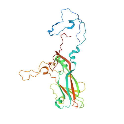
Coxsackievirus capsid protein VP1A [auth 1]305Coxsackievirus A24Mutation(s): 0 
UniProt
Find proteins for V9VEF3 (Coxsackievirus A24)
Explore V9VEF3 
Go to UniProtKB:  V9VEF3
Entity Groups  
Sequence Clusters30% Identity50% Identity70% Identity90% Identity95% Identity100% Identity
UniProt GroupV9VEF3
Sequence Annotations
Expand
  • Reference Sequence
Find similar proteins by:  (by identity cutoff)  |  3D Structure
Entity ID: 2
MoleculeChains Sequence LengthOrganismDetailsImage
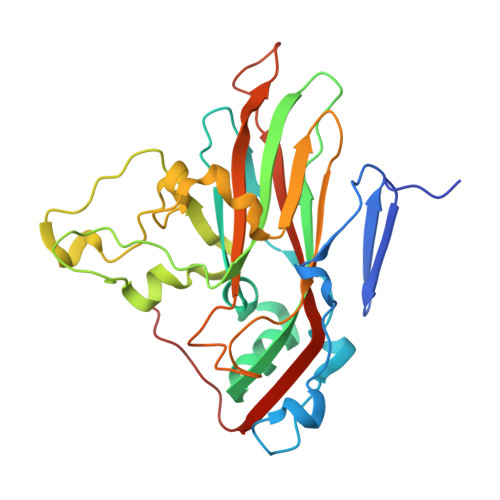
Coxsackievirus capsid protein VP2B [auth 2]271Coxsackievirus A24Mutation(s): 0 
UniProt
Find proteins for V9VEF3 (Coxsackievirus A24)
Explore V9VEF3 
Go to UniProtKB:  V9VEF3
Entity Groups  
Sequence Clusters30% Identity50% Identity70% Identity90% Identity95% Identity100% Identity
UniProt GroupV9VEF3
Sequence Annotations
Expand
  • Reference Sequence
Find similar proteins by:  (by identity cutoff)  |  3D Structure
Entity ID: 3
MoleculeChains Sequence LengthOrganismDetailsImage
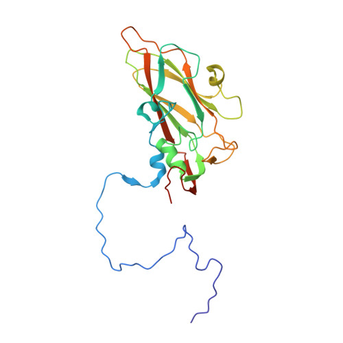
Coxsackievirus capsid protein VP3C [auth 3]240Coxsackievirus A24Mutation(s): 0 
UniProt
Find proteins for V9VEF3 (Coxsackievirus A24)
Explore V9VEF3 
Go to UniProtKB:  V9VEF3
Entity Groups  
Sequence Clusters30% Identity50% Identity70% Identity90% Identity95% Identity100% Identity
UniProt GroupV9VEF3
Sequence Annotations
Expand
  • Reference Sequence
Find similar proteins by:  (by identity cutoff)  |  3D Structure
Entity ID: 4
MoleculeChains Sequence LengthOrganismDetailsImage
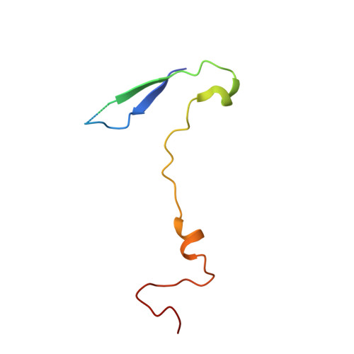
Coxsackievirus capsid protein VP4D [auth 4]69Coxsackievirus A24Mutation(s): 0 
UniProt
Find proteins for V9VEF3 (Coxsackievirus A24)
Explore V9VEF3 
Go to UniProtKB:  V9VEF3
Entity Groups  
Sequence Clusters30% Identity50% Identity70% Identity90% Identity95% Identity100% Identity
UniProt GroupV9VEF3
Sequence Annotations
Expand
  • Reference Sequence
Small Molecules
Ligands 5 Unique
IDChains Name / Formula / InChI Key2D Diagram3D Interactions
SIA
Query on SIA

Download Ideal Coordinates CCD File 
E [auth 1]N-acetyl-alpha-neuraminic acid
C11 H19 N O9
SQVRNKJHWKZAKO-YRMXFSIDSA-N
HEZ
Query on HEZ

Download Ideal Coordinates CCD File 
F [auth 1],
G [auth 1]
HEXANE-1,6-DIOL
C6 H14 O2
XXMIOPMDWAUFGU-UHFFFAOYSA-N
CA
Query on CA

Download Ideal Coordinates CCD File 
I [auth 1],
J [auth 1],
K [auth 1],
O [auth 2],
Q [auth 3]
CALCIUM ION
Ca
BHPQYMZQTOCNFJ-UHFFFAOYSA-N
CL
Query on CL

Download Ideal Coordinates CCD File 
L [auth 1],
M [auth 1],
N [auth 1],
P [auth 2],
R [auth 3]
CHLORIDE ION
Cl
VEXZGXHMUGYJMC-UHFFFAOYSA-M
NA
Query on NA

Download Ideal Coordinates CCD File 
H [auth 1]SODIUM ION
Na
FKNQFGJONOIPTF-UHFFFAOYSA-N
Experimental Data & Validation

Experimental Data

  • Method: X-RAY DIFFRACTION
  • Resolution: 1.65 Å
  • R-Value Work: 0.149 
  • R-Value Observed: 0.149 
  • Space Group: I 2 2 2
Unit Cell:
Length ( Å )Angle ( ˚ )
a = 306.398α = 90
b = 366.096β = 90
c = 367.7γ = 90
Software Package:
Software NamePurpose
ISPyBdata collection
PYMOLmodel building
REFMACrefinement
XDSdata reduction
XSCALEdata scaling
PYMOLphasing

Structure Validation

View Full Validation Report


Ligand Structure Quality Assessment 


Entry History 

Deposition Data

Revision History  (Full details and data files)

  • Version 1.0: 2014-11-05
    Type: Initial release
  • Version 1.1: 2020-07-29
    Type: Remediation
    Reason: Carbohydrate remediation
    Changes: Data collection, Derived calculations, Structure summary
  • Version 1.2: 2024-02-28
    Changes: Data collection, Database references, Derived calculations, Structure summary