4PYH

Phospho-glucan bound structure of starch phosphatase Starch EXcess4 reveals the mechanism for C6-specificty


Experimental Data Snapshot

  • Method: X-RAY DIFFRACTION
  • Resolution: 1.65 Å
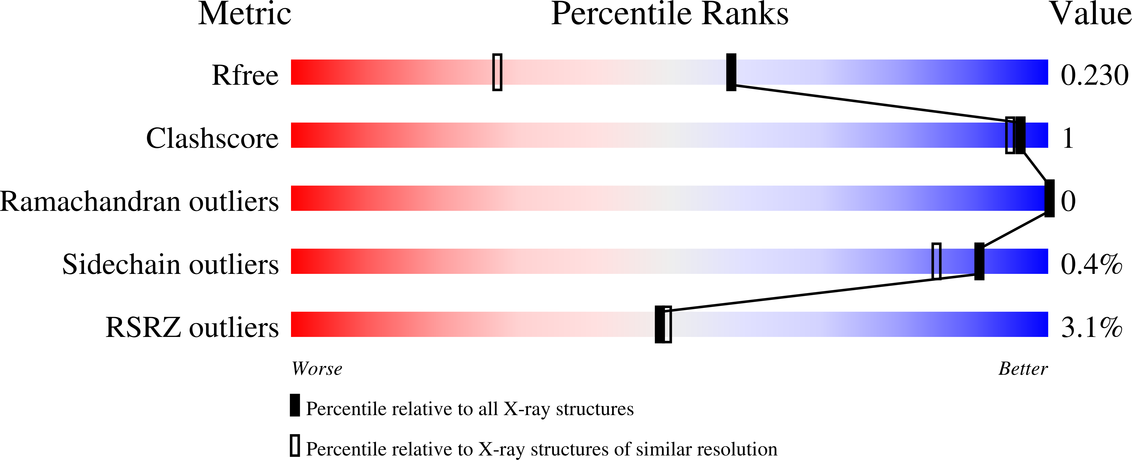
  • R-Value Free: 0.218 
  • R-Value Work: 0.177 
  • R-Value Observed: 0.179 

Starting Model: experimental
View more details

wwPDB Validation   3D Report Full Report


This is version 2.1 of the entry. See complete history


Literature

Phosphoglucan-bound structure of starch phosphatase Starch Excess4 reveals the mechanism for C6 specificity.

Meekins, D.A.Raththagala, M.Husodo, S.White, C.J.Guo, H.F.Kotting, O.Vander Kooi, C.W.Gentry, M.S.

(2014) Proc Natl Acad Sci U S A 111: 7272-7277

  • DOI: https://doi.org/10.1073/pnas.1400757111
  • Primary Citation of Related Structures:  
    4PYH

  • PubMed Abstract: 

    Plants use the insoluble polyglucan starch as their primary glucose storage molecule. Reversible phosphorylation, at the C6 and C3 positions of glucose moieties, is the only known natural modification of starch and is the key regulatory mechanism controlling its diurnal breakdown in plant leaves. The glucan phosphatase Starch Excess4 (SEX4) is a position-specific starch phosphatase that is essential for reversible starch phosphorylation; its absence leads to a dramatic accumulation of starch in Arabidopsis, but the basis for its function is unknown. Here we describe the crystal structure of SEX4 bound to maltoheptaose and phosphate to a resolution of 1.65 Å. SEX4 binds maltoheptaose via a continuous binding pocket and active site that spans both the carbohydrate-binding module (CBM) and the dual-specificity phosphatase (DSP) domain. This extended interface is composed of aromatic and hydrophilic residues that form a specific glucan-interacting platform. SEX4 contains a uniquely adapted DSP active site that accommodates a glucan polymer and is responsible for positioning maltoheptaose in a C6-specific orientation. We identified two DSP domain residues that are responsible for SEX4 site-specific activity and, using these insights, we engineered a SEX4 double mutant that completely reversed specificity from the C6 to the C3 position. Our data demonstrate that the two domains act in consort, with the CBM primarily responsible for engaging glucan chains, whereas the DSP integrates them in the catalytic site for position-specific dephosphorylation. These data provide important insights into the structural basis of glucan phosphatase site-specific activity and open new avenues for their biotechnological utilization.


  • Organizational Affiliation

    Department of Molecular and Cellular Biochemistry and Center for Structural Biology, University of Kentucky, Lexington, KY 40535-0509; and.


Macromolecules
Find similar proteins by:  (by identity cutoff)  |  3D Structure
Entity ID: 1
MoleculeChains Sequence LengthOrganismDetailsImage
Phosphoglucan phosphatase DSP4, chloroplastic294Arabidopsis thalianaMutation(s): 1 
Gene Names: DSP4PTPKIS1SEX4At3g52180F4F15.290
EC: 3.1.3
UniProt
Find proteins for Q9FEB5 (Arabidopsis thaliana)
Explore Q9FEB5 
Go to UniProtKB:  Q9FEB5
Entity Groups  
Sequence Clusters30% Identity50% Identity70% Identity90% Identity95% Identity100% Identity
UniProt GroupQ9FEB5
Sequence Annotations
Expand
  • Reference Sequence
Oligosaccharides

Help

Entity ID: 2
MoleculeChains Length2D Diagram Glycosylation3D Interactions
alpha-D-glucopyranose-(1-4)-alpha-D-glucopyranose-(1-4)-alpha-D-glucopyranose-(1-4)-alpha-D-glucopyranose-(1-4)-alpha-D-glucopyranose-(1-4)-alpha-D-glucopyranose
B
6N/A
Glycosylation Resources
GlyTouCan:  G91368RN
GlyCosmos:  G91368RN
GlyGen:  G91368RN
Biologically Interesting Molecules (External Reference) 1 Unique
Experimental Data & Validation

Experimental Data

  • Method: X-RAY DIFFRACTION
  • Resolution: 1.65 Å
  • R-Value Free: 0.218 
  • R-Value Work: 0.177 
  • R-Value Observed: 0.179 
  • Space Group: P 21 21 21
Unit Cell:
Length ( Å )Angle ( ˚ )
a = 33.475α = 90
b = 77.893β = 90
c = 117.213γ = 90
Software Package:
Software NamePurpose
HKL-2000data collection
PHENIXmodel building
REFMACrefinement
HKL-2000data reduction
HKL-2000data scaling
PHENIXphasing

Structure Validation

View Full Validation Report


Entry History 

Deposition Data

Revision History  (Full details and data files)

  • Version 1.0: 2014-04-23
    Type: Initial release
  • Version 1.1: 2014-06-18
    Changes: Database references
  • Version 2.0: 2020-07-29
    Type: Remediation
    Reason: Carbohydrate remediation
    Changes: Atomic model, Data collection, Database references, Derived calculations, Structure summary
  • Version 2.1: 2023-09-20
    Changes: Data collection, Database references, Refinement description, Structure summary