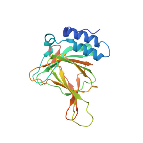
4PJY

Azide bound Cysteine Dioxygenase at pH 6.2

  • Classification: OXIDOREDUCTASE
  • Organism(s): Rattus norvegicus
  • Expression System: Escherichia coli
  • Mutation(s): No 

  • Deposited: 2014-05-12 Released: 2016-02-17 
  • Deposition Author(s): Driggers, C.M., Karplus, P.A.
  • Funding Organization(s): National Institutes of Health/National Institute of Diabetes and Digestive and Kidney Disease (NIH/NIDDK), Department of Energy (DOE, United States)

Experimental Data Snapshot

  • Method: X-RAY DIFFRACTION
  • Resolution: 1.50 Å
  • R-Value Free: 0.211 
  • R-Value Work: 0.174 
  • R-Value Observed: 0.178 

wwPDB Validation   3D Report Full Report


This is version 1.5 of the entry. See complete history


Literature

Structure-Based Insights into the Role of the Cys-Tyr Crosslink and Inhibitor Recognition by Mammalian Cysteine Dioxygenase.

Driggers, C.M.Kean, K.M.Hirschberger, L.L.Cooley, R.B.Stipanuk, M.H.Karplus, P.A.

(2016) J Mol Biol 428: 3999-4012

  • DOI: https://doi.org/10.1016/j.jmb.2016.07.012
  • Primary Citation of Related Structures:  
    4PIX, 4PIY, 4PIZ, 4PJY, 4XET, 4XEZ, 4XF0, 4XF1, 4XF3, 4XF4, 4XF9, 4XFB, 4XFC, 4XFF, 4XFG, 4XFH, 4XFI, 5I0R, 5I0S, 5I0T, 5I0U

  • PubMed Abstract: 

    In mammals, the non-heme iron enzyme cysteine dioxygenase (CDO) helps regulate Cys levels through converting Cys to Cys sulfinic acid. Its activity is in part modulated by the formation of a Cys93-Tyr157 crosslink that increases its catalytic efficiency over 10-fold. Here, 21 high-resolution mammalian CDO structures are used to gain insight into how the Cys-Tyr crosslink promotes activity and how select competitive inhibitors bind. Crystal structures of crosslink-deficient C93A and Y157F variants reveal similar ~1.0-Å shifts in the side chain of residue 157, and both variant structures have a new chloride ion coordinating the active site iron. Cys binding is also different from wild-type CDO, and no Cys-persulfenate forms in the C93A or Y157F active sites at pH6.2 or 8.0. We conclude that the crosslink enhances activity by positioning the Tyr157 hydroxyl to enable proper Cys binding, proper oxygen binding, and optimal chemistry. In addition, structures are presented for homocysteine (Hcy), D-Cys, thiosulfate, and azide bound as competitive inhibitors. The observed binding modes of Hcy and D-Cys clarify why they are not substrates, and the binding of azide shows that in contrast to what has been proposed, it does not bind in these crystals as a superoxide mimic.


  • Organizational Affiliation

    Department of Biochemistry and Biophysics, 2011 Ag & Life Sciences Building, Oregon State University, Corvallis, OR 97331, USA.


Macromolecules
Find similar proteins by:  (by identity cutoff)  |  3D Structure
Entity ID: 1
MoleculeChains Sequence LengthOrganismDetailsImage
Cysteine dioxygenase type 1200Rattus norvegicusMutation(s): 0 
Gene Names: Cdo1
EC: 1.13.11.20
UniProt
Find proteins for P21816 (Rattus norvegicus)
Explore P21816 
Go to UniProtKB:  P21816
Entity Groups  
Sequence Clusters30% Identity50% Identity70% Identity90% Identity95% Identity100% Identity
UniProt GroupP21816
Sequence Annotations
Expand
  • Reference Sequence
Experimental Data & Validation

Experimental Data

  • Method: X-RAY DIFFRACTION
  • Resolution: 1.50 Å
  • R-Value Free: 0.211 
  • R-Value Work: 0.174 
  • R-Value Observed: 0.178 
  • Space Group: P 43 21 2
Unit Cell:
Length ( Å )Angle ( ˚ )
a = 57.6α = 90
b = 57.6β = 90
c = 122.4γ = 90
Software Package:
Software NamePurpose
PHENIXrefinement

Structure Validation

View Full Validation Report


Entry History & Funding Information

Deposition Data


Funding OrganizationLocationGrant Number
National Institutes of Health/National Institute of Diabetes and Digestive and Kidney Disease (NIH/NIDDK)United StatesDK-056649
Department of Energy (DOE, United States)United StatesDE-AC02-05CH11231

Revision History  (Full details and data files)

  • Version 1.0: 2016-02-17
    Type: Initial release
  • Version 1.1: 2016-12-14
    Changes: Database references
  • Version 1.2: 2017-09-20
    Changes: Author supporting evidence
  • Version 1.3: 2019-12-04
    Changes: Author supporting evidence
  • Version 1.4: 2023-12-27
    Changes: Data collection, Database references, Derived calculations
  • Version 1.5: 2024-10-23
    Changes: Structure summary