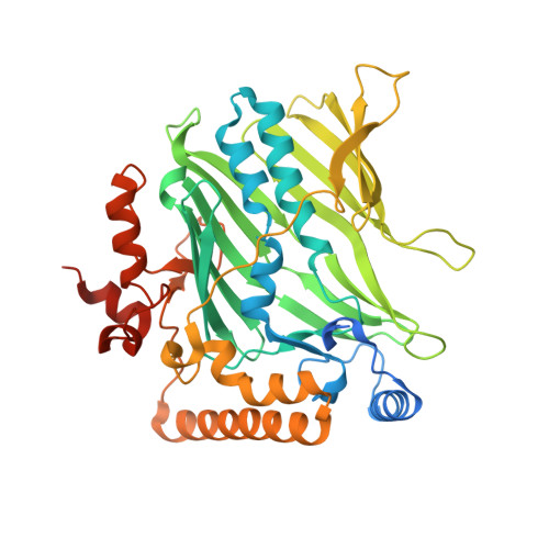
4PH7

Structure of Osh6p in complex with phosphatidylinositol 4-phosphate


Experimental Data Snapshot

  • Method: X-RAY DIFFRACTION
  • Resolution: 2.55 Å
  • R-Value Free: 0.237 
  • R-Value Work: 0.188 
  • R-Value Observed: 0.191 

Starting Model: experimental
View more details

wwPDB Validation   3D Report Full Report


Ligand Structure Quality Assessment 


This is version 2.2 of the entry. See complete history


Literature

INTRACELLULAR TRANSPORT. Phosphatidylserine transport by ORP/Osh proteins is driven by phosphatidylinositol 4-phosphate.

Moser von Filseck, J.Copic, A.Delfosse, V.Vanni, S.Jackson, C.L.Bourguet, W.Drin, G.

(2015) Science 349: 432-436

  • DOI: https://doi.org/10.1126/science.aab1346
  • Primary Citation of Related Structures:  
    4PH7

  • PubMed Abstract: 

    In eukaryotic cells, phosphatidylserine (PS) is synthesized in the endoplasmic reticulum (ER) but is highly enriched in the plasma membrane (PM), where it contributes negative charge and to specific recruitment of signaling proteins. This distribution relies on transport mechanisms whose nature remains elusive. Here, we found that the PS transporter Osh6p extracted phosphatidylinositol 4-phosphate (PI4P) and exchanged PS for PI4P between two membranes. We solved the crystal structure of Osh6p:PI4P complex and demonstrated that the transport of PS by Osh6p depends on PI4P recognition in vivo. Finally, we showed that the PI4P-phosphatase Sac1p, by maintaining a PI4P gradient at the ER/PM interface, drove PS transport. Thus, PS transport by oxysterol-binding protein-related protein (ORP)/oxysterol-binding homology (Osh) proteins is fueled by PI4P metabolism through PS/PI4P exchange cycles.


  • Organizational Affiliation

    Institut de Pharmacologie Moléculaire et Cellulaire, Université de Nice Sophia-Antipolis and CNRS, 660 route des lucioles, 06560 Valbonne, France.


Macromolecules
Find similar proteins by:  (by identity cutoff)  |  3D Structure
Entity ID: 1
MoleculeChains Sequence LengthOrganismDetailsImage
Oxysterol-binding protein homolog 6
A, B, C, D
450Saccharomyces cerevisiae S288CMutation(s): 0 
Gene Names: OSH6YKR003WYK102
Membrane Entity: Yes 
UniProt
Find proteins for Q02201 (Saccharomyces cerevisiae (strain ATCC 204508 / S288c))
Explore Q02201 
Go to UniProtKB:  Q02201
Entity Groups  
Sequence Clusters30% Identity50% Identity70% Identity90% Identity95% Identity100% Identity
UniProt GroupQ02201
Sequence Annotations
Expand
  • Reference Sequence
Small Molecules
Ligands 1 Unique
IDChains Name / Formula / InChI Key2D Diagram3D Interactions
2Y5
Query on 2Y5

Download Ideal Coordinates CCD File 
E [auth A],
F [auth B],
G [auth C],
H [auth D]
(2R)-1-{[(R)-hydroxy{[(1R,2R,3R,4R,5S,6R)-2,3,5,6-tetrahydroxy-4-(phosphonooxy)cyclohexyl]oxy}phosphoryl]oxy}-3-(octadecanoyloxy)propan-2-yl (5Z,8Z,11Z,14Z)-icosa-5,8,11,14-tetraenoate
C47 H84 O16 P2
ROKMWIUSHIHOGI-BWTMTLBLSA-N
Experimental Data & Validation

Experimental Data

  • Method: X-RAY DIFFRACTION
  • Resolution: 2.55 Å
  • R-Value Free: 0.237 
  • R-Value Work: 0.188 
  • R-Value Observed: 0.191 
  • Space Group: P 21 21 21
Unit Cell:
Length ( Å )Angle ( ˚ )
a = 114.11α = 90
b = 122.26β = 90
c = 141.49γ = 90
Software Package:
Software NamePurpose
PHENIXrefinement
XDSdata reduction
XSCALEdata scaling
PHENIXphasing

Structure Validation

View Full Validation Report


Ligand Structure Quality Assessment 


Entry History & Funding Information

Deposition Data


Funding OrganizationLocationGrant Number
French National Research AgencyFrance2010-1503-01
ERC programAdvanced Grant 268888

Revision History  (Full details and data files)

  • Version 1.0: 2015-07-29
    Type: Initial release
  • Version 1.1: 2015-08-05
    Changes: Database references
  • Version 2.0: 2017-09-06
    Changes: Advisory, Atomic model, Author supporting evidence, Derived calculations
  • Version 2.1: 2017-12-27
    Changes: Structure summary
  • Version 2.2: 2023-12-20
    Changes: Data collection, Database references, Refinement description, Structure summary