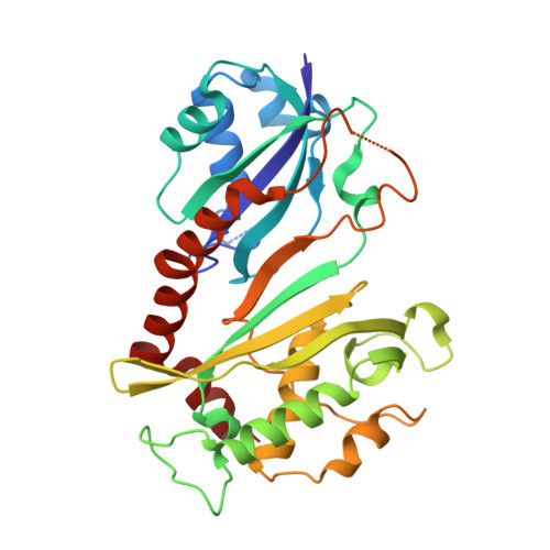
4NZ7

Steroid receptor RNA Activator (SRA) modification by the human Pseudouridine Synthase 1 (hPus1p): RNA binding, activity, and atomic model


Experimental Data Snapshot

  • Method: X-RAY DIFFRACTION
  • Resolution: 2.70 Å
  • R-Value Free: 0.238 
  • R-Value Work: 0.183 
  • R-Value Observed: 0.189 

wwPDB Validation   3D Report Full Report


This is version 1.1 of the entry. See complete history


Literature

Steroid Receptor RNA Activator (SRA) Modification by the Human Pseudouridine Synthase 1 (hPus1p): RNA Binding, Activity, and Atomic Model

Huet, T.Miannay, F.-A.Patton, J.R.Thore, S.

(2014) PLoS One 9: e94610-e94610

  • DOI: https://doi.org/10.1371/journal.pone.0094610
  • Primary Citation of Related Structures:  
    4NZ6, 4NZ7

  • PubMed Abstract: 

    The most abundant of the modified nucleosides, and once considered as the "fifth" nucleotide in RNA, is pseudouridine, which results from the action of pseudouridine synthases. Recently, the mammalian pseudouridine synthase 1 (hPus1p) has been reported to modulate class I and class II nuclear receptor responses through its ability to modify the Steroid receptor RNA Activator (SRA). These findings highlight a new level of regulation in nuclear receptor (NR)-mediated transcriptional responses. We have characterised the RNA association and activity of the human Pus1p enzyme with its unusual SRA substrate. We validate that the minimal RNA fragment within SRA, named H7, is necessary for both the association and modification by hPus1p. Furthermore, we have determined the crystal structure of the catalytic domain of hPus1p at 2.0 Å resolution, alone and in a complex with several molecules present during crystallisation. This model shows an extended C-terminal helix specifically found in the eukaryotic protein, which may prevent the enzyme from forming a homodimer, both in the crystal lattice and in solution. Our biochemical and structural data help to understand the hPus1p active site architecture, and detail its particular requirements with regard to one of its nuclear substrates, the non-coding RNA SRA.


  • Organizational Affiliation

    Department of Molecular Biology, University of Geneva, Sciences III, Geneva, Switzerland.


Macromolecules
Find similar proteins by:  (by identity cutoff)  |  3D Structure
Entity ID: 1
MoleculeChains Sequence LengthOrganismDetailsImage
tRNA pseudouridine synthase A, mitochondrial313Homo sapiensMutation(s): 1 
Gene Names: PUS1PP8985
EC: 5.4.99.12 (PDB Primary Data), 5.4.99 (UniProt)
UniProt & NIH Common Fund Data Resources
Find proteins for Q9Y606 (Homo sapiens)
Explore Q9Y606 
Go to UniProtKB:  Q9Y606
PHAROS:  Q9Y606
GTEx:  ENSG00000177192 
Entity Groups  
Sequence Clusters30% Identity50% Identity70% Identity90% Identity95% Identity100% Identity
UniProt GroupQ9Y606
Sequence Annotations
Expand
  • Reference Sequence
Small Molecules
Ligands 2 Unique
IDChains Name / Formula / InChI Key2D Diagram3D Interactions
EPE
Query on EPE

Download Ideal Coordinates CCD File 
B [auth A]4-(2-HYDROXYETHYL)-1-PIPERAZINE ETHANESULFONIC ACID
C8 H18 N2 O4 S
JKMHFZQWWAIEOD-UHFFFAOYSA-N
PGE
Query on PGE

Download Ideal Coordinates CCD File 
C [auth A]TRIETHYLENE GLYCOL
C6 H14 O4
ZIBGPFATKBEMQZ-UHFFFAOYSA-N
Experimental Data & Validation

Experimental Data

  • Method: X-RAY DIFFRACTION
  • Resolution: 2.70 Å
  • R-Value Free: 0.238 
  • R-Value Work: 0.183 
  • R-Value Observed: 0.189 
  • Space Group: P 2 21 21
Unit Cell:
Length ( Å )Angle ( ˚ )
a = 39.446α = 90
b = 69.044β = 90
c = 116.797γ = 90
Software Package:
Software NamePurpose
DNAdata collection
PHASERphasing
PHENIXrefinement
XDSdata reduction
XSCALEdata scaling

Structure Validation

View Full Validation Report


Entry History 

Deposition Data

Revision History  (Full details and data files)

  • Version 1.0: 2014-05-21
    Type: Initial release
  • Version 1.1: 2024-03-20
    Changes: Data collection, Database references, Derived calculations