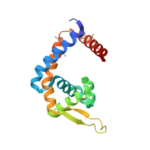
The molecular mechanisms of allosteric mutations impairing MepR repressor function in multidrug-resistant strains of Staphylococcus aureus.
Birukou, I., Tonthat, N.K., Seo, S.M., Schindler, B.D., Kaatz, G.W., Brennan, R.G.(2013) mBio 4: e00528-e00513
- PubMed: 23982071
- DOI: https://doi.org/10.1128/mBio.00528-13
- Primary Citation of Related Structures:
4L9J, 4L9N, 4L9T, 4L9V, 4LD5 - PubMed Abstract:
Overexpression of the Staphylococcus aureus multidrug efflux pump MepA confers resistance to a wide variety of antimicrobials. mepA expression is controlled by MarR family member MepR, which represses mepA and autorepresses its own production. Mutations in mepR are a primary cause of mepA overexpression in clinical isolates of multidrug-resistant S. aureus. Here, we report crystal structures of three multidrug-resistant MepR variants, which contain the single-amino-acid substitution A103V, F27L, or Q18P, and wild-type MepR in its DNA-bound conformation. Although each mutation impairs MepR function by decreasing its DNA binding affinity, none is located in the DNA binding domain. Rather, all are found in the linker region connecting the dimerization and DNA binding domains. Specifically, the A103V substitution impinges on F27, which resolves potential steric clashes via displacement of the DNA binding winged-helix-turn-helix motifs that lead to a 27-fold reduction in DNA binding affinity. The F27L substitution forces F104 into an alternative rotamer, which kinks helix 5, thereby interfering with the positioning of the DNA binding domains and decreasing mepR operator affinity by 35-fold. The Q18P mutation affects the MepR structure and function most significantly by either creating kinks in the middle of helix 1 or completely unfolding its C terminus. In addition, helix 5 of Q18P is either bent or completely dissected into two smaller helices. Consequently, DNA binding is diminished by 2,000-fold. Our structural studies reveal heretofore-unobserved allosteric mechanisms that affect repressor function of a MarR family member and result in multidrug-resistant Staphylococcus aureus. Staphylococcus aureus is a major health threat to immunocompromised patients. S. aureus multidrug-resistant variants that overexpress the multidrug efflux pump mepA emerge frequently due to point mutations in MarR family member MepR, the mepA transcription repressor. Significantly, the majority of MepR mutations identified in these S. aureus clinical isolates are found not in the DNA binding domain but rather in a linker region, connecting the dimerization and DNA binding domains. The location of these mutants underscores the critical importance of a properly functioning allosteric mechanism that regulates MepR function. Understanding the dysregulation of such allosteric MepR mutants underlies this study. The high-resolution structures of three such allosteric MepR mutants reveal unpredictable conformational consequences, all of which preclude cognate DNA binding, while biochemical studies emphasize their debilitating effects on DNA binding affinity. Hence, mutations in the linker region of MepR and their structural consequences are key generators of multidrug-resistant Staphylococcus aureus.
Organizational Affiliation:
Department of Biochemistry, Duke University School of Medicine, Durham, North Carolina, USA.


