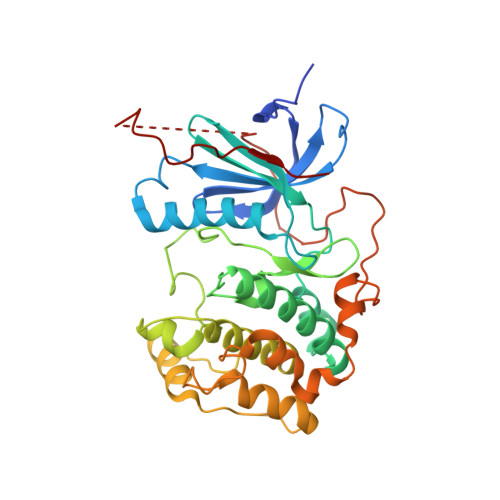
Crystal structures of S6K1 provide insights into the regulation mechanism of S6K1 by the hydrophobic motif
Wang, J., Zhong, C., Wang, F., Qu, F., Ding, J.(2013) Biochem J 454: 39-47
- PubMed: 23731517
- DOI: https://doi.org/10.1042/BJ20121863
- Primary Citation of Related Structures:
4L3J, 4L3L, 4L42, 4L43, 4L44, 4L45, 4L46 - PubMed Abstract:
The activity of S6K1 (p70 ribosomal protein subunit 6 kinase 1) is stimulated by phosphorylation of Thr389 in the hydrophobic motif by mTORC1 (mammalian target of rapamycin complex 1) and phosphorylation of Thr229 in the activation loop by PDK1 (phosphoinositide-dependent kinase 1); however, the order of the two events is still ambiguous. In the present paper we report six crystal structures of the S6K1 kinase domain alone or plus the hydrophobic motif in various forms, in complexes with a highly specific inhibitor. The structural data, together with the biochemical data, reveal in vivo phosphorylation of Thr389 in the absence of Thr229 phosphorylation and demonstrate the importance of two conserved residues, Gln140 and Arg121, in the establishment of a hydrogen-bonding network between the N-lobe (N-terminal lobe) and the hydrophobic motif. Phosphorylation of Thr389 or introduction of a corresponding negatively charged group leads to reinforcement of the network and stabilization of helix αC. Furthermore, comparisons of S6K1 with other AGC (protein kinase A/protein kinase G/protein kinase C) family kinases suggest that the structural and sequence differences in the hydrophobic motif and helix αC account for their divergence in PDK1 dependency. Taken together, the results of the present study indicate that phosphorylation of the hydrophobic motif in S6K1 is independent of, and probably precedes and promotes, phosphorylation of the activation loop.
Organizational Affiliation:
State Key Laboratory of Molecular Biology, Institute of Biochemistry and Cell Biology, Shanghai Institute for Biological Sciences, Chinese Academy of Sciences, 320 Yue-Yang Road, Shanghai 200031, China.


