4KNL

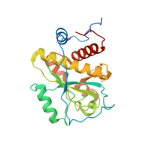
Crystal structure of Staphylococcus aureus hydrolase AmiA in complex with its ligand


Experimental Data Snapshot

  • Method: X-RAY DIFFRACTION
  • Resolution: 1.55 Å
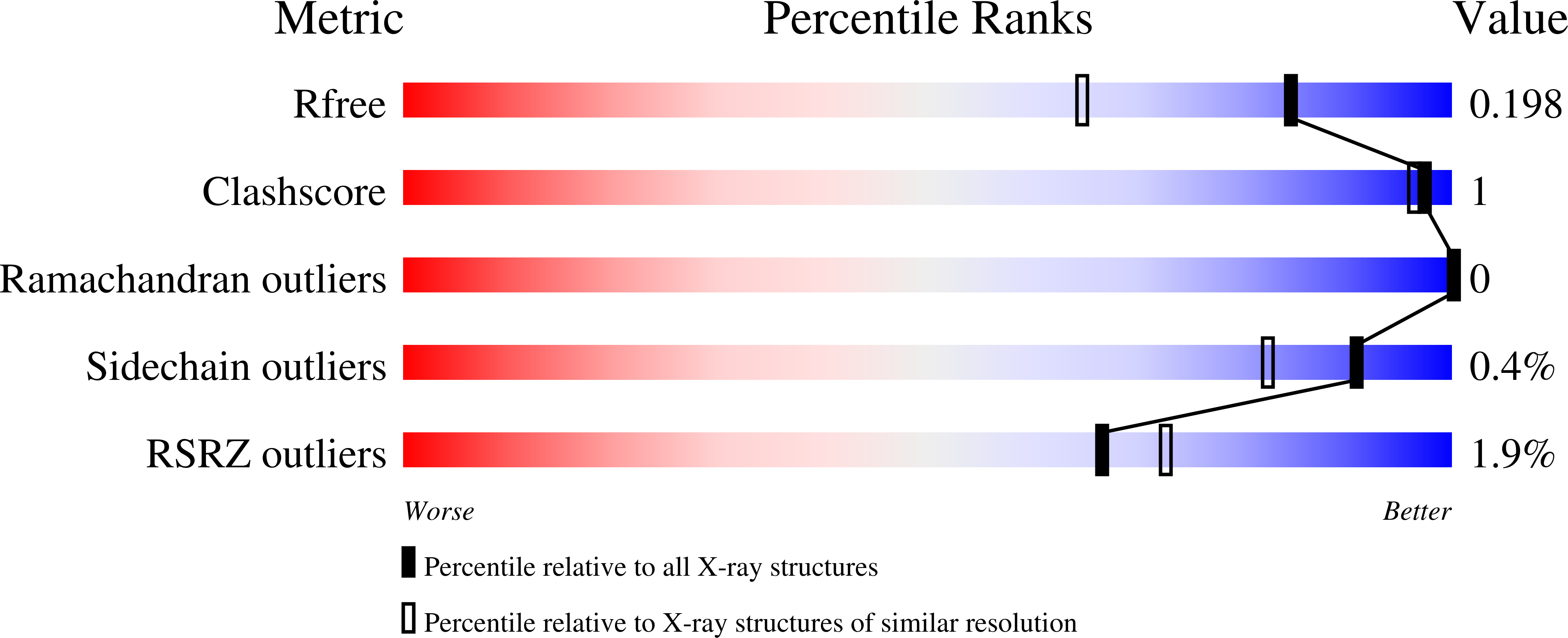
  • R-Value Free: 0.198 
  • R-Value Work: 0.171 
  • R-Value Observed: 0.172 

Starting Model: experimental
View more details

wwPDB Validation   3D Report Full Report


Ligand Structure Quality Assessment 


This is version 3.0 of the entry. See complete history


Literature

Structure-function analysis of Staphylococcus aureus amidase reveals the determinants of peptidoglycan recognition and cleavage.

Buttner, F.M.Zoll, S.Nega, M.Gotz, F.Stehle, T.

(2014) J Biol Chem 289: 11083-11094

  • DOI: https://doi.org/10.1074/jbc.M114.557306
  • Primary Citation of Related Structures:  
    4KNK, 4KNL

  • PubMed Abstract: 

    The bifunctional major autolysin AtlA of Staphylococcus aureus cleaves the bacterium's peptidoglycan network (PGN) at two distinct sites during cell division. Deletion of the enzyme results in large cell clusters with disordered division patterns, indicating that AtlA could be a promising target for the development of new antibiotics. One of the two functions of AtlA is performed by the N-acetylmuramyl-l-alanine amidase AmiA, which cleaves the bond between the carbohydrate and the peptide moieties of PGN. To establish the structural requirements of PGN recognition and the enzymatic mechanism of cleavage, we solved the crystal structure of the catalytic domain of AmiA (AmiA-cat) in complex with a peptidoglycan-derived ligand at 1.55 Å resolution. The peptide stem is clearly visible in the structure, forming extensive contacts with protein residues by docking into an elongated groove. Less well defined electron density and the analysis of surface features indicate likely positions of the carbohydrate backbone and the pentaglycine bridge. Substrate specificity analysis supports the importance of the pentaglycine bridge for fitting into the binding cleft of AmiA-cat. PGN of S. aureus with l-lysine tethered with d-alanine via a pentaglycine bridge is completely hydrolyzed, whereas PGN of Bacillus subtilis with meso-diaminopimelic acid directly tethered with d-alanine is not hydrolyzed. An active site mutant, H370A, of AmiA-cat was completely inactive, providing further support for the proposed catalytic mechanism of AmiA. The structure reported here is not only the first of any bacterial amidase in which both the PGN component and the water molecule that carries out the nucleophilic attack on the carbonyl carbon of the scissile bond are present; it is also the first peptidoglycan amidase complex structure of an important human pathogen.


  • Organizational Affiliation

    Interfaculty Institute of Biochemistry, University of Tübingen, Hoppe-Seyler-Strasse 4, 72076 Tübingen, Germany.


Macromolecules
Find similar proteins by:  (by identity cutoff)  |  3D Structure
Entity ID: 1
MoleculeChains Sequence LengthOrganismDetailsImage
Bifunctional autolysin
A, B, C, D
225Staphylococcus aureus subsp. aureus NCTC 8325Mutation(s): 0 
Gene Names: atlnagSAOUHSC_00994
EC: 3.5.1.28 (PDB Primary Data), 3.2.1.96 (PDB Primary Data)
UniProt
Find proteins for Q2FZK7 (Staphylococcus aureus (strain NCTC 8325 / PS 47))
Explore Q2FZK7 
Go to UniProtKB:  Q2FZK7
Entity Groups  
Sequence Clusters30% Identity50% Identity70% Identity90% Identity95% Identity100% Identity
UniProt GroupQ2FZK7
Sequence Annotations
Expand
  • Reference Sequence

Find similar proteins by:  Sequence   |   3D Structure  

Entity ID: 2
MoleculeChains Sequence LengthOrganismDetailsImage
Muramyl tetrapeptideE [auth F],
F [auth G]
5synthetic constructMutation(s): 0 
Sequence Annotations
Expand
  • Reference Sequence
Small Molecules
Ligands 7 Unique
IDChains Name / Formula / InChI Key2D Diagram3D Interactions
MUB
Query on MUB

Download Ideal Coordinates CCD File 
X [auth F],
Y [auth G]
N-acetyl-alpha-muramic acid
C11 H19 N O8
MNLRQHMNZILYPY-MDMHTWEWSA-N
TAR
Query on TAR

Download Ideal Coordinates CCD File 
S [auth C]D(-)-TARTARIC ACID
C4 H6 O6
FEWJPZIEWOKRBE-LWMBPPNESA-N
MPD
Query on MPD

Download Ideal Coordinates CCD File 
R [auth C](4S)-2-METHYL-2,4-PENTANEDIOL
C6 H14 O2
SVTBMSDMJJWYQN-YFKPBYRVSA-N
OXM
Query on OXM

Download Ideal Coordinates CCD File 
O [auth B]OXAMIC ACID
C2 H3 N O3
SOWBFZRMHSNYGE-UHFFFAOYSA-N
IMD
Query on IMD

Download Ideal Coordinates CCD File 
G [auth A],
K [auth B],
P [auth C],
U [auth D]
IMIDAZOLE
C3 H5 N2
RAXXELZNTBOGNW-UHFFFAOYSA-O
FMT
Query on FMT

Download Ideal Coordinates CCD File 
I [auth A],
M [auth B]
FORMIC ACID
C H2 O2
BDAGIHXWWSANSR-UHFFFAOYSA-N
NA
Query on NA

Download Ideal Coordinates CCD File 
H [auth A]
J [auth A]
L [auth B]
N [auth B]
Q [auth C]
H [auth A],
J [auth A],
L [auth B],
N [auth B],
Q [auth C],
T [auth C],
V [auth D],
W [auth D]
SODIUM ION
Na
FKNQFGJONOIPTF-UHFFFAOYSA-N
Modified Residues  1 Unique
IDChains TypeFormula2D DiagramParent
ALY
Query on ALY
E [auth F],
F [auth G]
L-PEPTIDE LINKINGC8 H16 N2 O3LYS
Biologically Interesting Molecules (External Reference) 1 Unique
Entity ID: 2
IDChains NameType/Class2D Diagram3D Interactions
PRD_001066
Query on PRD_001066
E [auth F],
F [auth G]
Muramyl tetrapeptidePeptide-like / Unknown
Experimental Data & Validation

Experimental Data

  • Method: X-RAY DIFFRACTION
  • Resolution: 1.55 Å
  • R-Value Free: 0.198 
  • R-Value Work: 0.171 
  • R-Value Observed: 0.172 
  • Space Group: P 1 21 1
Unit Cell:
Length ( Å )Angle ( ˚ )
a = 67.81α = 90
b = 82.83β = 96.18
c = 77.43γ = 90
Software Package:
Software NamePurpose
RemDAqdata collection
PHASERphasing
PHENIXrefinement
XDSdata reduction
XSCALEdata scaling

Structure Validation

View Full Validation Report


Ligand Structure Quality Assessment 


Entry History 

Deposition Data

Revision History  (Full details and data files)

  • Version 1.0: 2014-03-12
    Type: Initial release
  • Version 1.1: 2014-12-03
    Changes: Database references
  • Version 1.2: 2020-07-29
    Type: Remediation
    Reason: Carbohydrate remediation
    Changes: Advisory, Data collection, Database references, Derived calculations, Structure summary
  • Version 2.0: 2023-03-01
    Changes: Atomic model, Data collection, Database references, Derived calculations, Polymer sequence, Source and taxonomy, Structure summary
  • Version 2.1: 2023-09-20
    Changes: Data collection, Refinement description
  • Version 3.0: 2023-11-15
    Changes: Atomic model, Data collection, Derived calculations