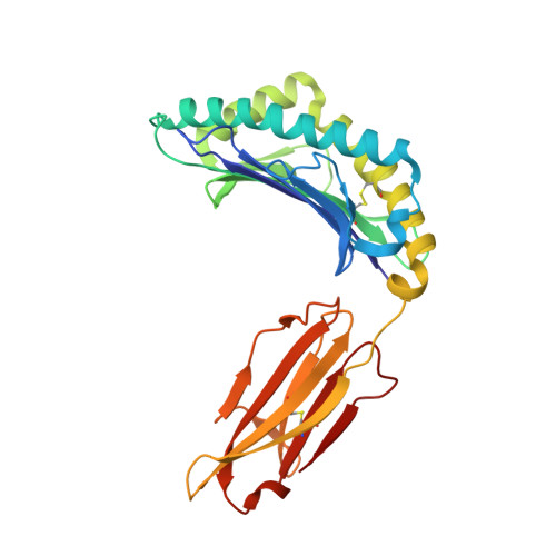
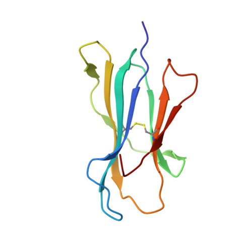
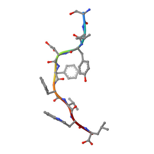
4HX1

Structure of HLA-A68 complexed with a tumor antigen derived peptide


Experimental Data Snapshot

  • Method: X-RAY DIFFRACTION
  • Resolution: 1.80 Å
  • R-Value Free: 0.226 
  • R-Value Work: 0.190 
  • R-Value Observed: 0.192 

wwPDB Validation   3D Report Full Report


This is version 1.0 of the entry. See complete history


Literature

Structural basis for the differential classification of HLA-A*6802 and HLA-A*6801 into the A2 and A3 supertypes

Niu, L.Cheng, H.Zhang, S.Tan, S.Zhang, Y.Qi, J.Liu, J.Gao, G.F.

(2013) Mol Immunol 55: 381-392

  • DOI: https://doi.org/10.1016/j.molimm.2013.03.015
  • Primary Citation of Related Structures:  
    4HWZ, 4HX1, 4I48

  • PubMed Abstract: 

    High polymorphism is one of the most important features of human leukocyte antigen (HLA) alleles, which were initially classified by serotyping but have recently been re-grouped into supertypes according to their peptide presentation properties. Two relatively prevalent HLA alleles HLA-A*6801 and HLA-A*6802, are classified into the same serotype HLA-A68. However, based on their distinct peptide-binding characteristics, HLA-A*6801 is grouped into A3 supertype, whereas HLA-A*6802 belongs to A2 supertype, similar to HLA-A*0201. Thusfar, the structural basis of the different supertype definitions of these serotyping-identical HLA alleles remains largely unknown. Herein, we determined the structures of HLA-A*6801 and HLA-A*6802 presenting three typical A3 and A2 supertype-restricted peptides, respectively. The binding capabilities of these peptides to HLA-A*6801, HLA-A*6802, and HLA-A*0201 were analyzed. These data indicate that the similar conformations of the residues within the F pocket contribute to close-related peptide binding features of HLA-A*6802 and HLA-A*0201. However, the overall structure and the peptide conformation of HLA-A*6802 are more similar to HLA-A*6801 rather than HLA-A*0201 which illuminates the similar serotype grouping of HLA-A*6802 and HLA-A*6801. Our findings are helpful for understanding the divergent peptide presentation and virus-specific CTL responses impacted by MHC micropolymorphisms and also elucidate the molecular basis of HLA supertype definitions.


  • Organizational Affiliation

    CAS Key Laboratory of Pathogenic Microbiology and Immunology, Institute of Microbiology, Chinese Academy of Sciences, Beijing, China.


Macromolecules
Find similar proteins by:  (by identity cutoff)  |  3D Structure
Entity ID: 1
MoleculeChains Sequence LengthOrganismDetailsImage
MHC class I antigen274Homo sapiensMutation(s): 0 
Gene Names: HLA-A
UniProt & NIH Common Fund Data Resources
Find proteins for P04439 (Homo sapiens)
Explore P04439 
Go to UniProtKB:  P04439
PHAROS:  P04439
GTEx:  ENSG00000206503 
Entity Groups  
Sequence Clusters30% Identity50% Identity70% Identity90% Identity95% Identity100% Identity
UniProt GroupP04439
Sequence Annotations
Expand
  • Reference Sequence
Find similar proteins by:  (by identity cutoff)  |  3D Structure
Entity ID: 2
MoleculeChains Sequence LengthOrganismDetailsImage
Beta-2-microglobulin99Homo sapiensMutation(s): 0 
Gene Names: B2M
UniProt & NIH Common Fund Data Resources
Find proteins for P61769 (Homo sapiens)
Explore P61769 
Go to UniProtKB:  P61769
PHAROS:  P61769
GTEx:  ENSG00000166710 
Entity Groups  
Sequence Clusters30% Identity50% Identity70% Identity90% Identity95% Identity100% Identity
UniProt GroupP61769
Sequence Annotations
Expand
  • Reference Sequence

Find similar proteins by:  Sequence   |   3D Structure  

Entity ID: 3
MoleculeChains Sequence LengthOrganismDetailsImage
9-mer peptide from Tyrosinase-related protein-29Homo sapiensMutation(s): 0 
EC: 5.3.3.12
UniProt & NIH Common Fund Data Resources
Find proteins for P40126 (Homo sapiens)
Explore P40126 
Go to UniProtKB:  P40126
PHAROS:  P40126
GTEx:  ENSG00000080166 
Entity Groups  
UniProt GroupP40126
Sequence Annotations
Expand
  • Reference Sequence
Small Molecules
Ligands 1 Unique
IDChains Name / Formula / InChI Key2D Diagram3D Interactions
GOL
Query on GOL

Download Ideal Coordinates CCD File 
D [auth A]GLYCEROL
C3 H8 O3
PEDCQBHIVMGVHV-UHFFFAOYSA-N
Experimental Data & Validation

Experimental Data

  • Method: X-RAY DIFFRACTION
  • Resolution: 1.80 Å
  • R-Value Free: 0.226 
  • R-Value Work: 0.190 
  • R-Value Observed: 0.192 
  • Space Group: C 1 2 1
Unit Cell:
Length ( Å )Angle ( ˚ )
a = 129.237α = 90
b = 37.857β = 120.04
c = 108.323γ = 90
Software Package:
Software NamePurpose
HKL-2000data collection
CNSrefinement
PHENIXrefinement
HKL-2000data reduction
HKL-2000data scaling
CNSphasing

Structure Validation

View Full Validation Report


Entry History 

Deposition Data

Revision History  (Full details and data files)

  • Version 1.0: 2013-10-02
    Type: Initial release