X-ray crystallographic studies of family 11 xylanase Michaelis and product complexes: implications for the catalytic mechanism.
Wan, Q., Zhang, Q., Hamilton-Brehm, S., Weiss, K., Mustyakimov, M., Coates, L., Langan, P., Graham, D., Kovalevsky, A.(2014) Acta Crystallogr D Biol Crystallogr 70: 11-23
- PubMed: 24419374
- DOI: https://doi.org/10.1107/S1399004713023626
- Primary Citation of Related Structures:
4HK8, 4HK9, 4HKL, 4HKO, 4HKW, 6K9X - PubMed Abstract:
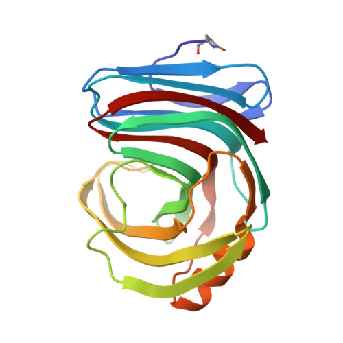
Xylanases catalyze the hydrolysis of plant hemicellulose xylan into oligosaccharides by cleaving the main-chain glycosidic linkages connecting xylose subunits. To study ligand binding and to understand how the pH constrains the activity of the enzyme, variants of the Trichoderma reesei xylanase were designed to either abolish its activity (E177Q) or to change its pH optimum (N44H). An E177Q-xylohexaose complex structure was obtained at 1.15 Å resolution which represents a pseudo-Michaelis complex and confirmed the conformational movement of the thumb region owing to ligand binding. Co-crystallization of N44H with xylohexaose resulted in a hydrolyzed xylotriose bound in the active site. Co-crystallization of the wild-type enzyme with xylopentaose trapped an aglycone xylotriose and a transglycosylated glycone product. Replacing amino acids near Glu177 decreased the xylanase activity but increased the relative activity at alkaline pH. The substrate distortion in the E177Q-xylohexaose structure expands the possible conformational itinerary of this xylose ring during the enzyme-catalyzed xylan-hydrolysis reaction.
Organizational Affiliation:
Biology and Soft Matter Division, Oak Ridge National Laboratory, PO Box 2008, Oak Ridge, TN 37831, USA.


