4G01

ARA7-GDP-Ca2+/VPS9a


Experimental Data Snapshot

  • Method: X-RAY DIFFRACTION
  • Resolution: 2.20 Å
  • R-Value Free: 0.273 
  • R-Value Work: 0.198 
  • R-Value Observed: 0.202 

Starting Model: experimental
View more details

wwPDB Validation   3D Report Full Report


Ligand Structure Quality Assessment 


This is version 1.2 of the entry. See complete history


Literature

Direct metal recognition by guanine nucleotide-exchange factor in the initial step of the exchange reaction

Uejima, T.Ihara, K.Sunada, M.Kawasaki, M.Ueda, T.Kato, R.Nakano, A.Wakatsuki, S.

(2013) Acta Crystallogr D Biol Crystallogr 69: 345-351

  • DOI: https://doi.org/10.1107/S0907444912047294
  • Primary Citation of Related Structures:  
    4G01

  • PubMed Abstract: 

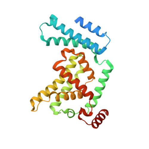
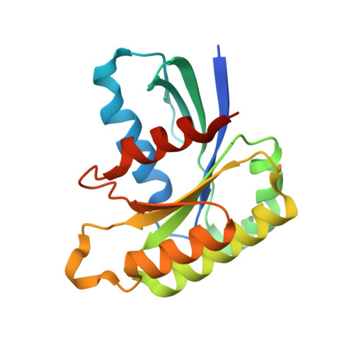
    Rab small GTPases regulate vesicle transport in eukaryotes by interacting with various effectors. Guanine nucleotide-exchange factor (GEF) catalyzes the transition from inactive GDP-bound Rab to active GTP-bound Rab. The existence of several GDP-bound intermediates containing the Arabidopsis thaliana Rab5 homologue ARA7 and the GEF VPS9a prior to the formation of a nucleotide-free binary complex has been proposed [Uejima et al. (2010), J. Biol. Chem. 285, 36689-36697]. During this process, VPS9a directly interacts with the β-phosphate of GDP and the P-loop lysine of ARA7 via a catalytically important aspartate finger, which promotes the release of GDP from ARA7. However, it is unclear how VPS9a removes Mg2+ from ARA7 before forming the GDP-bound ternary complex. Here, the structure of the ARA7-GDP-Ca2+-VPS9a complex is reported, in which the aspartate finger directly coordinates the divalent metal ion. Ca2+ is bound to the canonical Mg2+-binding site, coordinated by the β-phosphate of GDP and the P-loop serine of ARA7. Unexpectedly, Ca2+ is further coordinated by the aspartate finger and the main chain of VPS9a. This structure may represent the earliest intermediate step in the GEF-catalyzed nucleotide-exchange reaction of ARA7 before the metal-free GDP-bound intermediates are created.


  • Organizational Affiliation

    Structural Biology Research Center, Institute of Materials Structure Science, High Energy Accelerator Research Organization, 1-1 Oho, Tsukuba, Ibaraki 305-0801, Japan. tamami.uejima@kek.jp


Macromolecules
Find similar proteins by:  (by identity cutoff)  |  3D Structure
Entity ID: 1
MoleculeChains Sequence LengthOrganismDetailsImage
Vacuolar protein sorting-associated protein 9A267Arabidopsis thalianaMutation(s): 0 
Gene Names: VPS9A
UniProt
Find proteins for Q9LT31 (Arabidopsis thaliana)
Explore Q9LT31 
Go to UniProtKB:  Q9LT31
Entity Groups  
Sequence Clusters30% Identity50% Identity70% Identity90% Identity95% Identity100% Identity
UniProt GroupQ9LT31
Sequence Annotations
Expand
  • Reference Sequence
Find similar proteins by:  (by identity cutoff)  |  3D Structure
Entity ID: 2
MoleculeChains Sequence LengthOrganismDetailsImage
Ras-related protein RABF2b181Arabidopsis thalianaMutation(s): 0 
Gene Names: ARA-7
UniProt
Find proteins for Q9SN68 (Arabidopsis thaliana)
Explore Q9SN68 
Go to UniProtKB:  Q9SN68
Entity Groups  
Sequence Clusters30% Identity50% Identity70% Identity90% Identity95% Identity100% Identity
UniProt GroupQ9SN68
Sequence Annotations
Expand
  • Reference Sequence
Small Molecules
Ligands 2 Unique
IDChains Name / Formula / InChI Key2D Diagram3D Interactions
GDP
Query on GDP

Download Ideal Coordinates CCD File 
D [auth B]GUANOSINE-5'-DIPHOSPHATE
C10 H15 N5 O11 P2
QGWNDRXFNXRZMB-UUOKFMHZSA-N
CA
Query on CA

Download Ideal Coordinates CCD File 
C [auth A]CALCIUM ION
Ca
BHPQYMZQTOCNFJ-UHFFFAOYSA-N
Experimental Data & Validation

Experimental Data

  • Method: X-RAY DIFFRACTION
  • Resolution: 2.20 Å
  • R-Value Free: 0.273 
  • R-Value Work: 0.198 
  • R-Value Observed: 0.202 
  • Space Group: P 21 21 21
Unit Cell:
Length ( Å )Angle ( ˚ )
a = 68.349α = 90
b = 76.139β = 90
c = 81.787γ = 90
Software Package:
Software NamePurpose
HKL-2000data collection
AMoREphasing
REFMACrefinement
HKL-2000data reduction
HKL-2000data scaling

Structure Validation

View Full Validation Report


Ligand Structure Quality Assessment 


Entry History 

Deposition Data

  • Released Date: 2013-02-27 
  • Deposition Author(s): Ihara, K.

Revision History  (Full details and data files)

  • Version 1.0: 2013-02-27
    Type: Initial release
  • Version 1.1: 2013-08-07
    Changes: Database references
  • Version 1.2: 2023-11-08
    Changes: Data collection, Database references, Derived calculations, Refinement description