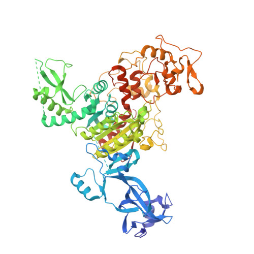
4FT4

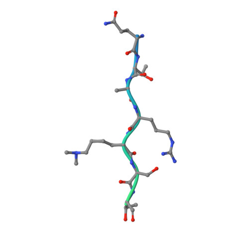
crystal structure of Zea mays ZMET2 in complex H3(1-32)K9me2 peptide and SAH


Experimental Data Snapshot

  • Method: X-RAY DIFFRACTION
  • Resolution: 2.70 Å
  • R-Value Free: 0.248 
  • R-Value Work: 0.206 
  • R-Value Observed: 0.208 

Starting Model: experimental
View more details

wwPDB Validation   3D Report Full Report


Ligand Structure Quality Assessment 


This is version 1.1 of the entry. See complete history


Literature

Dual Binding of Chromomethylase Domains to H3K9me2-Containing Nucleosomes Directs DNA Methylation in Plants.

Du, J.Zhong, X.Bernatavichute, Y.V.Stroud, H.Feng, S.Caro, E.Vashisht, A.A.Terragni, J.Chin, H.G.Tu, A.Hetzel, J.Wohlschlegel, J.A.Pradhan, S.Patel, D.J.Jacobsen, S.E.

(2012) Cell 151: 167-180

  • DOI: https://doi.org/10.1016/j.cell.2012.07.034
  • Primary Citation of Related Structures:  
    4FSX, 4FT2, 4FT4

  • PubMed Abstract: 

    DNA methylation and histone modification exert epigenetic control over gene expression. CHG methylation by CHROMOMETHYLASE3 (CMT3) depends on histone H3K9 dimethylation (H3K9me2), but the mechanism underlying this relationship is poorly understood. Here, we report multiple lines of evidence that CMT3 interacts with H3K9me2-containing nucleosomes. CMT3 genome locations nearly perfectly correlated with H3K9me2, and CMT3 stably associated with H3K9me2-containing nucleosomes. Crystal structures of maize CMT3 homolog ZMET2, in complex with H3K9me2 peptides, showed that ZMET2 binds H3K9me2 via both bromo adjacent homology (BAH) and chromo domains. The structures reveal an aromatic cage within both BAH and chromo domains as interaction interfaces that capture H3K9me2. Mutations that abolish either interaction disrupt CMT3 binding to nucleosomes and show a complete loss of CMT3 activity in vivo. Our study establishes dual recognition of H3K9me2 marks by BAH and chromo domains and reveals a distinct mechanism of interplay between DNA methylation and histone modification.


  • Organizational Affiliation

    Structural Biology Program, Memorial Sloan-Kettering Cancer Center, New York, NY 10065, USA.


Macromolecules
Find similar proteins by:  (by identity cutoff)  |  3D Structure
Entity ID: 1
MoleculeChains Sequence LengthOrganismDetailsImage
DNA (cytosine-5)-methyltransferase 1A [auth B],
B [auth A]
784Zea maysMutation(s): 0 
Gene Names: MET2AZMET2
EC: 2.1.1.37
UniProt
Find proteins for Q9AXT8 (Zea mays)
Explore Q9AXT8 
Go to UniProtKB:  Q9AXT8
Entity Groups  
Sequence Clusters30% Identity50% Identity70% Identity90% Identity95% Identity100% Identity
UniProt GroupQ9AXT8
Sequence Annotations
Expand
  • Reference Sequence
Find similar proteins by:  (by identity cutoff)  |  3D Structure
Entity ID: 2
MoleculeChains Sequence LengthOrganismDetailsImage
H3(1-32)K9me2 peptideC [auth P],
D [auth Q]
32N/AMutation(s): 0 
UniProt
Find proteins for P59226 (Arabidopsis thaliana)
Explore P59226 
Go to UniProtKB:  P59226
Entity Groups  
Sequence Clusters30% Identity50% Identity70% Identity90% Identity95% Identity100% Identity
UniProt GroupP59226
Sequence Annotations
Expand
  • Reference Sequence
Small Molecules
Modified Residues  1 Unique
IDChains TypeFormula2D DiagramParent
MLY
Query on MLY
C [auth P],
D [auth Q]
L-PEPTIDE LINKINGC8 H18 N2 O2LYS
Experimental Data & Validation

Experimental Data

  • Method: X-RAY DIFFRACTION
  • Resolution: 2.70 Å
  • R-Value Free: 0.248 
  • R-Value Work: 0.206 
  • R-Value Observed: 0.208 
  • Space Group: P 1 21 1
Unit Cell:
Length ( Å )Angle ( ˚ )
a = 64.645α = 90
b = 111.558β = 101.98
c = 151.48γ = 90
Software Package:
Software NamePurpose
ADSCdata collection
PHENIXmodel building
PHENIXrefinement
HKL-2000data reduction
HKL-2000data scaling
PHENIXphasing

Structure Validation

View Full Validation Report


Ligand Structure Quality Assessment 


Entry History 

Deposition Data

Revision History  (Full details and data files)

  • Version 1.0: 2012-10-17
    Type: Initial release
  • Version 1.1: 2023-09-13
    Changes: Data collection, Database references, Derived calculations, Refinement description