4F1Z

Crystal structures reveal the multi-ligand binding mechanism of the Staphylococcus aureus ClfB


Experimental Data Snapshot

  • Method: X-RAY DIFFRACTION
  • Resolution: 2.30 Å
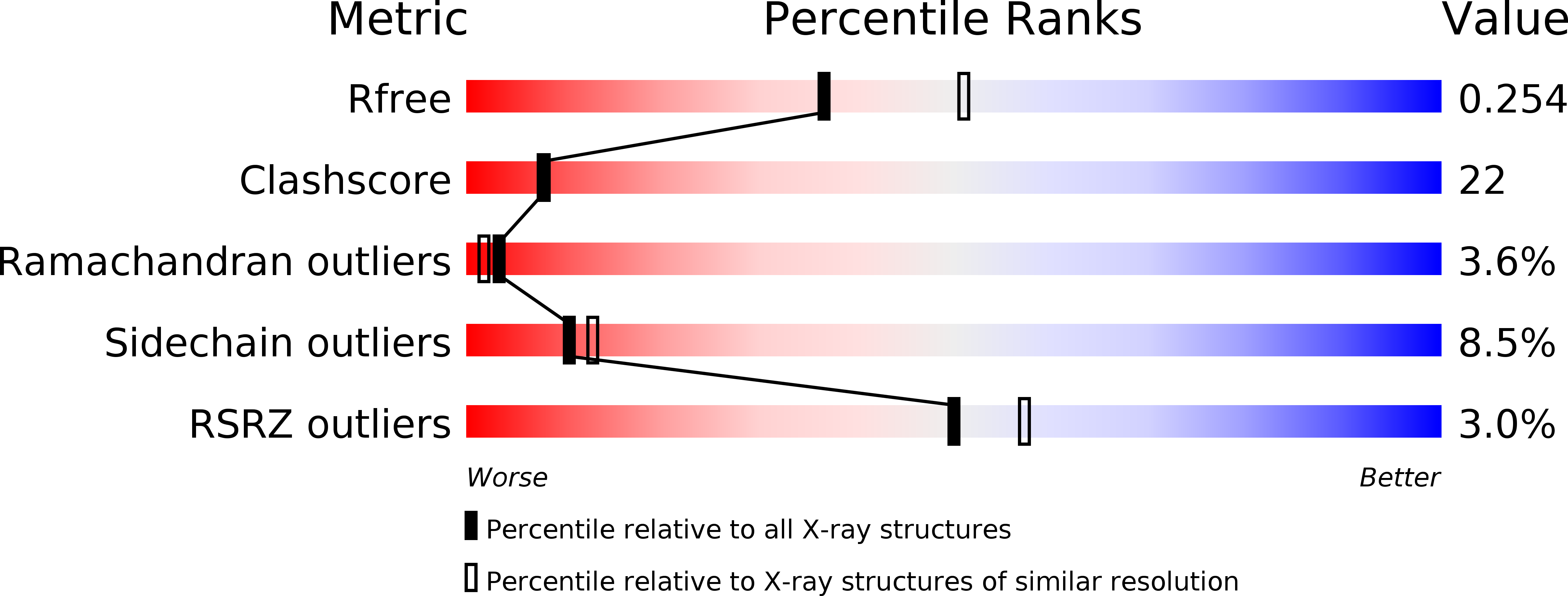
  • R-Value Free: 0.251 
  • R-Value Work: 0.247 
  • R-Value Observed: 0.248 

Starting Model: experimental
View more details

wwPDB Validation   3D Report Full Report


This is version 1.1 of the entry. See complete history


Literature

Crystal Structures Reveal the Multi-Ligand Binding Mechanism of Staphylococcus aureus ClfB

Xiang, H.Feng, Y.Wang, J.W.Liu, B.Chen, Y.G.Liu, L.Deng, X.M.Yang, M.J.

(2012) PLoS Pathog 8: e1002751-e1002751

  • DOI: https://doi.org/10.1371/journal.ppat.1002751
  • Primary Citation of Related Structures:  
    4F1Z, 4F20, 4F24, 4F27

  • PubMed Abstract: 

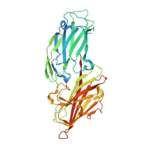
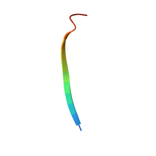
    Staphylococcus aureus (S. aureus) pathogenesis is a complex process involving a diverse array of extracellular and cell wall components. ClfB, an MSCRAMM (Microbial Surface Components Recognizing Adhesive Matrix Molecules) family surface protein, described as a fibrinogen-binding clumping factor, is a key determinant of S. aureus nasal colonization, but the molecular basis for ClfB-ligand recognition remains unknown. In this study, we solved the crystal structures of apo-ClfB and its complexes with fibrinogen α (Fg α) and cytokeratin 10 (CK10) peptides. Structural comparison revealed a conserved glycine-serine-rich (GSR) ClfB binding motif (GSSGXGXXG) within the ligands, which was also found in other human proteins such as Engrailed protein, TCF20 and Dermokine proteins. Interaction between Dermokine and ClfB was confirmed by subsequent binding assays. The crystal structure of ClfB complexed with a 15-residue peptide derived from Dermokine revealed the same peptide binding mode of ClfB as identified in the crystal structures of ClfB-Fg α and ClfB-CK10. The results presented here highlight the multi-ligand binding property of ClfB, which is very distinct from other characterized MSCRAMMs to-date. The adherence of multiple peptides carrying the GSR motif into the same pocket in ClfB is reminiscent of MHC molecules. Our results provide a template for the identification of other molecules targeted by S. aureus during its colonization and infection. We propose that other MSCRAMMs like ClfA and SdrG also possess multi-ligand binding properties.


  • Organizational Affiliation

    Key Laboratory for Protein Sciences of Ministry of Education, Tsinghua-Peking Center for Life Sciences, School of Life Sciences, Tsinghua University, Beijing, China.


Macromolecules
Find similar proteins by:  (by identity cutoff)  |  3D Structure
Entity ID: 1
MoleculeChains Sequence LengthOrganismDetailsImage
Clumping factor B363Staphylococcus aureus subsp. aureus MRSA252Mutation(s): 0 
Gene Names: clfBSAR2709
UniProt
Find proteins for Q6GDH2 (Staphylococcus aureus (strain MRSA252))
Explore Q6GDH2 
Go to UniProtKB:  Q6GDH2
Entity Groups  
Sequence Clusters30% Identity50% Identity70% Identity90% Identity95% Identity100% Identity
UniProt GroupQ6GDH2
Sequence Annotations
Expand
  • Reference Sequence

Find similar proteins by:  Sequence   |   3D Structure  

Entity ID: 2
MoleculeChains Sequence LengthOrganismDetailsImage
peptide from Keratin, type I cytoskeletal 10B [auth Q]14Homo sapiensMutation(s): 0 
UniProt & NIH Common Fund Data Resources
Find proteins for P13645 (Homo sapiens)
Explore P13645 
Go to UniProtKB:  P13645
PHAROS:  P13645
GTEx:  ENSG00000186395 
Entity Groups  
Sequence Clusters30% Identity50% Identity70% Identity90% Identity95% Identity100% Identity
UniProt GroupP13645
Sequence Annotations
Expand
  • Reference Sequence
Small Molecules
Ligands 1 Unique
IDChains Name / Formula / InChI Key2D Diagram3D Interactions
MG
Query on MG

Download Ideal Coordinates CCD File 
C [auth A]MAGNESIUM ION
Mg
JLVVSXFLKOJNIY-UHFFFAOYSA-N
Experimental Data & Validation

Experimental Data

  • Method: X-RAY DIFFRACTION
  • Resolution: 2.30 Å
  • R-Value Free: 0.251 
  • R-Value Work: 0.247 
  • R-Value Observed: 0.248 
  • Space Group: P 31 2 1
Unit Cell:
Length ( Å )Angle ( ˚ )
a = 70.331α = 90
b = 70.331β = 90
c = 177.157γ = 120
Software Package:
Software NamePurpose
HKL-2000data collection
PHASESphasing
PHENIXrefinement
HKL-2000data reduction
HKL-2000data scaling

Structure Validation

View Full Validation Report


Entry History 

Deposition Data

Revision History  (Full details and data files)

  • Version 1.0: 2012-08-08
    Type: Initial release
  • Version 1.1: 2023-11-08
    Changes: Data collection, Database references, Derived calculations, Refinement description