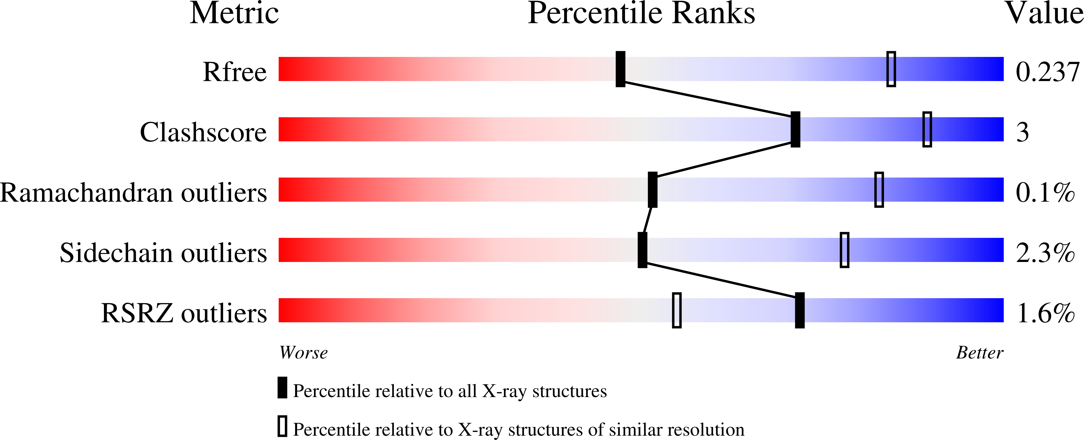
Ribosomal oxygenases are structurally conserved from prokaryotes to humans.
Chowdhury, R., Sekirnik, R., Brissett, N.C., Krojer, T., Ho, C.H., Ng, S.S., Clifton, I.J., Ge, W., Kershaw, N.J., Fox, G.C., Muniz, J.R.C., Vollmar, M., Phillips, C., Pilka, E.S., Kavanagh, K.L., von Delft, F., Oppermann, U., McDonough, M.A., Doherty, A.J., Schofield, C.J.(2014) Nature 510: 422-426
- PubMed: 24814345
- DOI: https://doi.org/10.1038/nature13263
- Primary Citation of Related Structures:
2XDV, 4BU2, 4BXF, 4CCJ, 4CCK, 4CCL, 4CCM, 4CCN, 4CCO, 4CSW, 4CUG, 4LIT, 4LIU, 4LIV - PubMed Abstract:
2-Oxoglutarate (2OG)-dependent oxygenases have important roles in the regulation of gene expression via demethylation of N-methylated chromatin components and in the hydroxylation of transcription factors and splicing factor proteins. Recently, 2OG-dependent oxygenases that catalyse hydroxylation of transfer RNA and ribosomal proteins have been shown to be important in translation relating to cellular growth, TH17-cell differentiation and translational accuracy. The finding that ribosomal oxygenases (ROXs) occur in organisms ranging from prokaryotes to humans raises questions as to their structural and evolutionary relationships. In Escherichia coli, YcfD catalyses arginine hydroxylation in the ribosomal protein L16; in humans, MYC-induced nuclear antigen (MINA53; also known as MINA) and nucleolar protein 66 (NO66) catalyse histidine hydroxylation in the ribosomal proteins RPL27A and RPL8, respectively. The functional assignments of ROXs open therapeutic possibilities via either ROX inhibition or targeting of differentially modified ribosomes. Despite differences in the residue and protein selectivities of prokaryotic and eukaryotic ROXs, comparison of the crystal structures of E. coli YcfD and Rhodothermus marinus YcfD with those of human MINA53 and NO66 reveals highly conserved folds and novel dimerization modes defining a new structural subfamily of 2OG-dependent oxygenases. ROX structures with and without their substrates support their functional assignments as hydroxylases but not demethylases, and reveal how the subfamily has evolved to catalyse the hydroxylation of different residue side chains of ribosomal proteins. Comparison of ROX crystal structures with those of other JmjC-domain-containing hydroxylases, including the hypoxia-inducible factor asparaginyl hydroxylase FIH and histone N(ε)-methyl lysine demethylases, identifies branch points in 2OG-dependent oxygenase evolution and distinguishes between JmjC-containing hydroxylases and demethylases catalysing modifications of translational and transcriptional machinery. The structures reveal that new protein hydroxylation activities can evolve by changing the coordination position from which the iron-bound substrate-oxidizing species reacts. This coordination flexibility has probably contributed to the evolution of the wide range of reactions catalysed by oxygenases.
Organizational Affiliation:
The Department of Chemistry and Oxford Centre for Integrative Systems Biology, University of Oxford, Mansfield Road, Oxford OX1 3TA, U.K.


