4CI3

Structure of the DDB1-CRBN E3 ubiquitin ligase bound to Pomalidomide


Experimental Data Snapshot

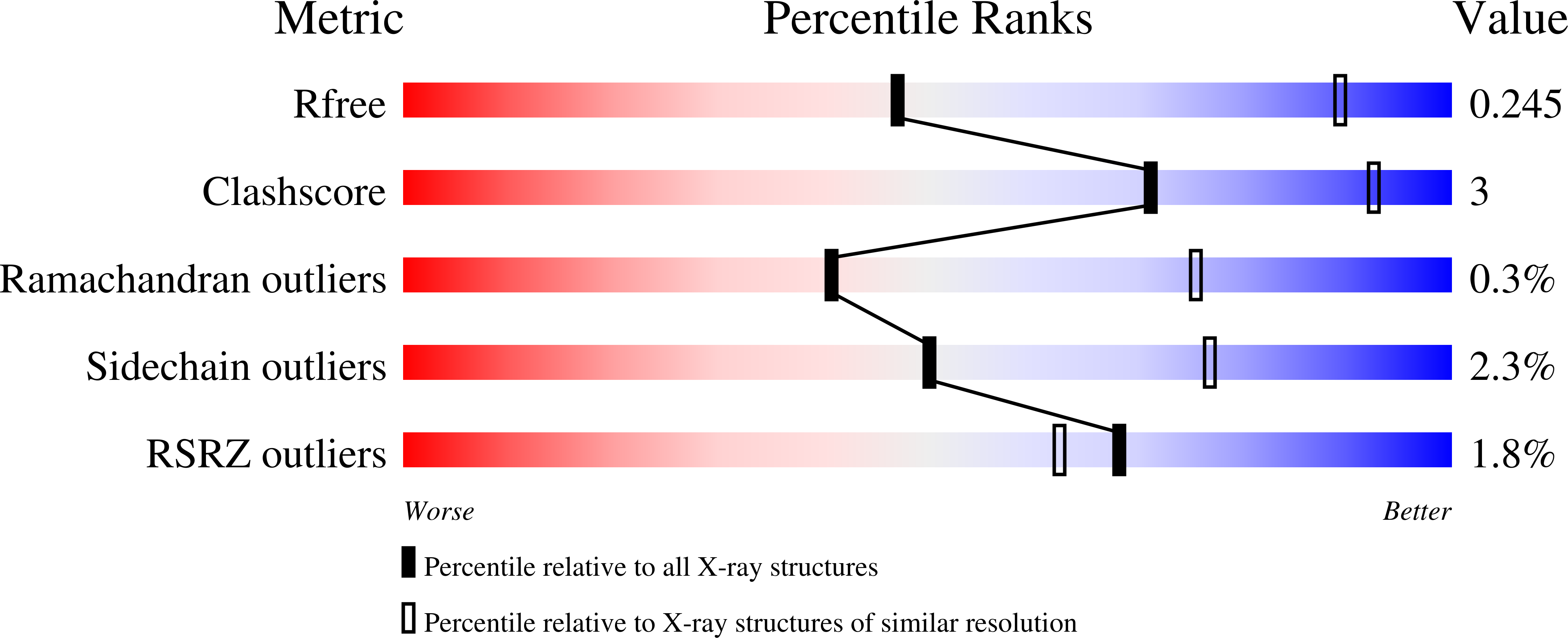
  • Method: X-RAY DIFFRACTION
  • Resolution: 3.50 Å
  • R-Value Free: 0.235 
  • R-Value Work: 0.211 
  • R-Value Observed: 0.212 

Starting Model: experimental
View more details

wwPDB Validation   3D Report Full Report


Ligand Structure Quality Assessment 


This is version 1.4 of the entry. See complete history


Literature

Structure of the Ddb1-Crbn E3 Ubiquitin Ligase in Complex with Thalidomide.

Fischer, E.S.Bohm, K.Lydeard, J.R.Yang, H.Stadler, M.B.Cavadini, S.Nagel, J.Serluca, F.Acker, V.Lingaraju, G.M.Tichkule, R.B.Schebesta, M.Forrester, W.C.Schirle, M.Hassiepen, U.Ottl, J.Hild, M.Beckwith, R.E.J.Harper, J.W.Jenkins, J.L.Thoma, N.H.

(2014) Nature 512: 49

  • DOI: https://doi.org/10.1038/nature13527
  • Primary Citation of Related Structures:  
    4CI1, 4CI2, 4CI3

  • PubMed Abstract: 

    In the 1950s, the drug thalidomide, administered as a sedative to pregnant women, led to the birth of thousands of children with multiple defects. Despite the teratogenicity of thalidomide and its derivatives lenalidomide and pomalidomide, these immunomodulatory drugs (IMiDs) recently emerged as effective treatments for multiple myeloma and 5q-deletion-associated dysplasia. IMiDs target the E3 ubiquitin ligase CUL4-RBX1-DDB1-CRBN (known as CRL4(CRBN)) and promote the ubiquitination of the IKAROS family transcription factors IKZF1 and IKZF3 by CRL4(CRBN). Here we present crystal structures of the DDB1-CRBN complex bound to thalidomide, lenalidomide and pomalidomide. The structure establishes that CRBN is a substrate receptor within CRL4(CRBN) and enantioselectively binds IMiDs. Using an unbiased screen, we identified the homeobox transcription factor MEIS2 as an endogenous substrate of CRL4(CRBN). Our studies suggest that IMiDs block endogenous substrates (MEIS2) from binding to CRL4(CRBN) while the ligase complex is recruiting IKZF1 or IKZF3 for degradation. This dual activity implies that small molecules can modulate an E3 ubiquitin ligase and thereby upregulate or downregulate the ubiquitination of proteins.


  • Organizational Affiliation

    1] Friedrich Miescher Institute for Biomedical Research, Maulbeerstrasse 66, CH-4058 Basel, Switzerland [2] University of Basel, Petersplatz 10, CH-4003 Basel, Switzerland.


Macromolecules
Find similar proteins by:  (by identity cutoff)  |  3D Structure
Entity ID: 1
MoleculeChains Sequence LengthOrganismDetailsImage
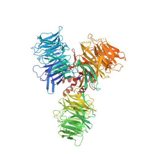
DNA DAMAGE-BINDING PROTEIN 11,158Homo sapiensMutation(s): 0 
UniProt & NIH Common Fund Data Resources
Find proteins for Q16531 (Homo sapiens)
Explore Q16531 
Go to UniProtKB:  Q16531
PHAROS:  Q16531
GTEx:  ENSG00000167986 
Entity Groups  
Sequence Clusters30% Identity50% Identity70% Identity90% Identity95% Identity100% Identity
UniProt GroupQ16531
Sequence Annotations
Expand
  • Reference Sequence
Find similar proteins by:  (by identity cutoff)  |  3D Structure
Entity ID: 2
MoleculeChains Sequence LengthOrganismDetailsImage
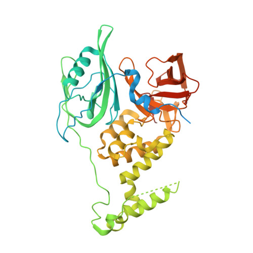
PROTEIN CEREBLON469Gallus gallusMutation(s): 0 
UniProt
Find proteins for P0CF65 (Gallus gallus)
Explore P0CF65 
Go to UniProtKB:  P0CF65
Entity Groups  
Sequence Clusters30% Identity50% Identity70% Identity90% Identity95% Identity100% Identity
UniProt GroupP0CF65
Sequence Annotations
Expand
  • Reference Sequence
Small Molecules
Ligands 2 Unique
IDChains Name / Formula / InChI Key2D Diagram3D Interactions
Y70
Query on Y70

Download Ideal Coordinates CCD File 
D [auth B]S-Pomalidomide
C13 H11 N3 O4
UVSMNLNDYGZFPF-QMMMGPOBSA-N
ZN
Query on ZN

Download Ideal Coordinates CCD File 
C [auth B]ZINC ION
Zn
PTFCDOFLOPIGGS-UHFFFAOYSA-N
Experimental Data & Validation

Experimental Data

  • Method: X-RAY DIFFRACTION
  • Resolution: 3.50 Å
  • R-Value Free: 0.235 
  • R-Value Work: 0.211 
  • R-Value Observed: 0.212 
  • Space Group: P 32 2 1
Unit Cell:
Length ( Å )Angle ( ˚ )
a = 171.07α = 90
b = 171.07β = 90
c = 137.93γ = 120
Software Package:
Software NamePurpose
BUSTERrefinement
XDSdata reduction
XSCALEdata scaling
PHASERphasing

Structure Validation

View Full Validation Report


Ligand Structure Quality Assessment 


Entry History 

Deposition Data

Revision History  (Full details and data files)

  • Version 1.0: 2014-07-16
    Type: Initial release
  • Version 1.1: 2014-07-30
    Changes: Database references
  • Version 1.2: 2014-08-13
    Changes: Database references
  • Version 1.3: 2019-03-27
    Changes: Data collection, Other, Source and taxonomy
  • Version 1.4: 2023-12-20
    Changes: Data collection, Database references, Derived calculations, Other, Refinement description