4C53

Crystal Structure of Guanarito virus GP2 in the post-fusion conformation


Experimental Data Snapshot

  • Method: X-RAY DIFFRACTION
  • Resolution: 4.14 Å
  • R-Value Free: 0.276 
  • R-Value Work: 0.255 
  • R-Value Observed: 0.256 

Starting Model: experimental
View more details

wwPDB Validation   3D Report Full Report


This is version 2.1 of the entry. See complete history


Literature

Crystal Structure of Venezuelan Hemorrhagic Fever Virus Fusion Glycoprotein Reveals a Class 1 Post-Fusion Architecture with Extensive Glycosylation.

Parsy, M.Harlos, K.Huiskonen, J.T.Bowden, T.A.

(2013) J Virol 87: 13070

  • DOI: https://doi.org/10.1128/JVI.02298-13
  • Primary Citation of Related Structures:  
    4C53

  • PubMed Abstract: 

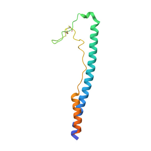
    Guanarito virus (GTOV) is an emergent and deadly pathogen. We present the crystal structure of the glycosylated GTOV fusion glycoprotein to 4.1-Å resolution in the postfusion conformation. Our structure reveals a classical six-helix bundle and presents direct verification that New World arenaviruses exhibit class I viral membrane fusion machinery. The structure provides visualization of an N-linked glycocalyx coat, and consideration of glycan dynamics reveals extensive coverage of the underlying protein surface, following virus-host membrane fusion.


  • Organizational Affiliation

    Division of Structural Biology, Wellcome Trust Centre for Human Genetics, University of Oxford, Oxford, United Kingdom.


Macromolecules
Find similar proteins by:  (by identity cutoff)  |  3D Structure
Entity ID: 1
MoleculeChains Sequence LengthOrganismDetailsImage
PRE-GLYCOPROTEIN POLYPROTEIN GP COMPLEX
A, B, C
138Mammarenavirus guanaritoenseMutation(s): 0 
UniProt
Find proteins for Q8AYW1 (Guanarito mammarenavirus (isolate Human/Venezuela/NH-95551/1990))
Explore Q8AYW1 
Go to UniProtKB:  Q8AYW1
Entity Groups  
Sequence Clusters30% Identity50% Identity70% Identity90% Identity95% Identity100% Identity
UniProt GroupQ8AYW1
Glycosylation
Glycosylation Sites: 3
Sequence Annotations
Expand
  • Reference Sequence
Oligosaccharides

Help

Entity ID: 2
MoleculeChains Length2D Diagram Glycosylation3D Interactions
beta-D-mannopyranose-(1-4)-2-acetamido-2-deoxy-beta-D-glucopyranose-(1-4)-2-acetamido-2-deoxy-beta-D-glucopyranose
D
3N-Glycosylation
Glycosylation Resources
GlyTouCan:  G15407YE
GlyCosmos:  G15407YE
GlyGen:  G15407YE
Entity ID: 3
MoleculeChains Length2D Diagram Glycosylation3D Interactions
2-acetamido-2-deoxy-beta-D-glucopyranose-(1-4)-2-acetamido-2-deoxy-beta-D-glucopyranose
E
2N-Glycosylation
Glycosylation Resources
GlyTouCan:  G42666HT
GlyCosmos:  G42666HT
GlyGen:  G42666HT
Entity ID: 4
MoleculeChains Length2D Diagram Glycosylation3D Interactions
alpha-D-mannopyranose-(1-3)-[alpha-D-mannopyranose-(1-6)]alpha-D-mannopyranose-(1-6)-[alpha-D-mannopyranose-(1-3)]beta-D-mannopyranose-(1-4)-2-acetamido-2-deoxy-beta-D-glucopyranose-(1-4)-2-acetamido-2-deoxy-beta-D-glucopyranose
F, G, H
7N-Glycosylation
Glycosylation Resources
GlyTouCan:  G55220VL
GlyCosmos:  G55220VL
GlyGen:  G55220VL
Experimental Data & Validation

Experimental Data

  • Method: X-RAY DIFFRACTION
  • Resolution: 4.14 Å
  • R-Value Free: 0.276 
  • R-Value Work: 0.255 
  • R-Value Observed: 0.256 
  • Space Group: P 41
Unit Cell:
Length ( Å )Angle ( ˚ )
a = 98.5α = 90
b = 98.5β = 90
c = 78.74γ = 90
Software Package:
Software NamePurpose
BUSTERrefinement
HKL-2000data reduction
SCALEPACKdata scaling
PHASERphasing

Structure Validation

View Full Validation Report


Entry History 

Deposition Data

Revision History  (Full details and data files)

  • Version 1.0: 2013-10-02
    Type: Initial release
  • Version 1.1: 2013-11-20
    Changes: Database references
  • Version 2.0: 2020-07-29
    Type: Remediation
    Reason: Carbohydrate remediation
    Changes: Atomic model, Data collection, Derived calculations, Other, Structure summary
  • Version 2.1: 2023-12-20
    Changes: Data collection, Database references, Refinement description, Structure summary