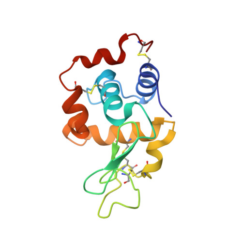
4BAF

Hen egg-white lysozyme structure in complex with the europium tris- hydroxyethyltriazoledipicolinate complex at 1.51 A resolution.


Experimental Data Snapshot

  • Method: X-RAY DIFFRACTION
  • Resolution: 1.51 Å
  • R-Value Free: 0.179 
  • R-Value Work: 0.156 
  • R-Value Observed: 0.158 

wwPDB Validation   3D Report Full Report


Ligand Structure Quality Assessment 


This is version 1.5 of the entry. See complete history


Literature

Clicked Europium Dipicolinate Complexes for Protein X-Ray Structure Determination.

Talon, R.Nauton, L.Canet, J.-L.Kahn, R.Girard, E.Gautier, A.

(2012) Chem Commun (Camb) 48: 11886

  • DOI: https://doi.org/10.1039/c2cc36982f
  • Primary Citation of Related Structures:  
    4BAD, 4BAF, 4BAL, 4BAP, 4BAR

  • PubMed Abstract: 

    New trisdipicolinic acid-lanthanide complexes are reported as phasing agents for X-ray crystallography of proteins. It is demonstrated that CuAAC modifications allow protein co-crystallization with low concentration of lanthanide complexes leading to an accurate structure determination.


  • Organizational Affiliation

    CEA, DSV, Institut de Biologie Structurale (IBS), 41 rue Jules Horowitz, Grenoble F-38027, France.


Macromolecules
Find similar proteins by:  (by identity cutoff)  |  3D Structure
Entity ID: 1
MoleculeChains Sequence LengthOrganismDetailsImage
LYSOZYME C129Gallus gallusMutation(s): 0 
EC: 3.2.1.17
UniProt
Find proteins for P00698 (Gallus gallus)
Explore P00698 
Go to UniProtKB:  P00698
Entity Groups  
Sequence Clusters30% Identity50% Identity70% Identity90% Identity95% Identity100% Identity
UniProt GroupP00698
Sequence Annotations
Expand
  • Reference Sequence
Small Molecules
Ligands 5 Unique
IDChains Name / Formula / InChI Key2D Diagram3D Interactions
IKX
Query on IKX

Download Ideal Coordinates CCD File 
C [auth A],
D [auth A],
E [auth A]
4-(4-(2-hydroxyethyl)-1H-1,2,3-triazol-1-yl)pyridine-2,6-dicarboxylic acid
C11 H12 N4 O5
RFMWOHVUEQDMAL-UHFFFAOYSA-N
EU3
Query on EU3

Download Ideal Coordinates CCD File 
B [auth A]EUROPIUM (III) ION
Eu
LNBHUCHAFZUEGJ-UHFFFAOYSA-N
ACT
Query on ACT

Download Ideal Coordinates CCD File 
F [auth A]ACETATE ION
C2 H3 O2
QTBSBXVTEAMEQO-UHFFFAOYSA-M
CL
Query on CL

Download Ideal Coordinates CCD File 
G [auth A]
H [auth A]
I [auth A]
J [auth A]
K [auth A]
G [auth A],
H [auth A],
I [auth A],
J [auth A],
K [auth A],
L [auth A],
O [auth A],
P [auth A],
Q [auth A]
CHLORIDE ION
Cl
VEXZGXHMUGYJMC-UHFFFAOYSA-M
NA
Query on NA

Download Ideal Coordinates CCD File 
M [auth A],
N [auth A]
SODIUM ION
Na
FKNQFGJONOIPTF-UHFFFAOYSA-N
Experimental Data & Validation

Experimental Data

  • Method: X-RAY DIFFRACTION
  • Resolution: 1.51 Å
  • R-Value Free: 0.179 
  • R-Value Work: 0.156 
  • R-Value Observed: 0.158 
  • Space Group: P 43 21 2
Unit Cell:
Length ( Å )Angle ( ˚ )
a = 77.763α = 90
b = 77.763β = 90
c = 37.896γ = 90
Software Package:
Software NamePurpose
PHENIXrefinement
XDSdata reduction
SCALAdata scaling
SHARPphasing

Structure Validation

View Full Validation Report


Ligand Structure Quality Assessment 


Entry History 

Deposition Data

Revision History  (Full details and data files)

  • Version 1.0: 2012-11-14
    Type: Initial release
  • Version 1.1: 2012-11-28
    Changes: Database references
  • Version 1.2: 2013-05-29
    Changes: Derived calculations
  • Version 1.3: 2015-04-01
    Changes: Database references
  • Version 1.4: 2019-03-06
    Changes: Data collection, Experimental preparation, Other
  • Version 1.5: 2024-10-16
    Changes: Data collection, Database references, Derived calculations, Other, Structure summary