4B56

Structure of ectonucleotide pyrophosphatase-phosphodiesterase-1 (NPP1)


Experimental Data Snapshot

  • Method: X-RAY DIFFRACTION
  • Resolution: 3.00 Å
  • R-Value Free: 0.234 
  • R-Value Work: 0.200 
  • R-Value Observed: 0.202 

Starting Model: experimental
View more details

wwPDB Validation   3D Report Full Report


Ligand Structure Quality Assessment 


This is version 2.1 of the entry. See complete history


Literature

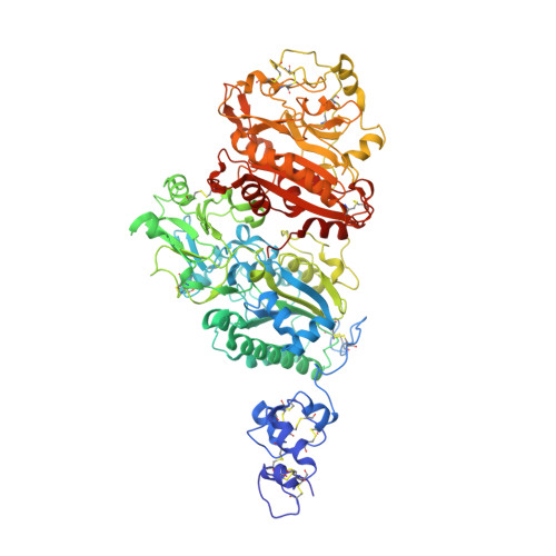
Structure of Npp1, an Ectonucleotide Pyrophosphatase/Phosphodiesterase Involved in Tissue Calcification.

Jansen, S.Perrakis, A.Ulens, C.Winkler, C.Andries, M.Joosten, R.P.Van Acker, M.Luyten, F.P.Moolenaar, W.H.Bollen, M.

(2012) Structure 20: 1948

  • DOI: https://doi.org/10.1016/j.str.2012.09.001
  • Primary Citation of Related Structures:  
    4B56

  • PubMed Abstract: 

    Ectonucleotide pyrophosphatase/phosphodiesterase-1 (NPP1) converts extracellular nucleotides into inorganic pyrophosphate, whereas its close relative NPP2/autotaxin hydrolyzes lysophospholipids. NPP1 regulates calcification in mineralization-competent tissues, and a lack of NPP1 function underlies calcification disorders. Here, we show that NPP1 forms homodimers via intramembrane disulfide bonding, but is also processed intracellularly to a secreted monomer. The structure of secreted NPP1 reveals a characteristic bimetallic active site and a nucleotide-binding groove, but it lacks the lipid-binding pocket and open tunnel present in NPP2. A loop adjacent to the nucleotide-binding site, which is disordered in NPP2, is well ordered in NPP1 and might promote nucleotide binding. Remarkably, the N-terminal somatomedin B-like domains of NPP1, unlike those in NPP2, are flexible and do not contact the catalytic domain. Our results provide a structural basis for the nucleotide pyrophosphatase activity of NPP1 and help to understand how disease-causing mutations may affect NPP1 structure and function.


  • Organizational Affiliation

    Laboratory of Biosignaling and Therapeutics, Department of Cellular and Molecular Medicine, University of Leuven, 3000 Leuven, Belgium.


Macromolecules
Find similar proteins by:  (by identity cutoff)  |  3D Structure
Entity ID: 1
MoleculeChains Sequence LengthOrganismDetailsImage
ECTONUCLEOTIDE PYROPHOSPHATASE/PHOSPHODIESTERASE FAMILY MEMBER 1
A, B
820Mus musculusMutation(s): 0 
EC: 3.1.4.1 (PDB Primary Data), 3.6.1.9 (PDB Primary Data)
UniProt & NIH Common Fund Data Resources
Find proteins for P06802 (Mus musculus)
Explore P06802 
Go to UniProtKB:  P06802
IMPC:  MGI:97370
Entity Groups  
Sequence Clusters30% Identity50% Identity70% Identity90% Identity95% Identity100% Identity
UniProt GroupP06802
Glycosylation
Glycosylation Sites: 4Go to GlyGen: P06802-1
Sequence Annotations
Expand
  • Reference Sequence
Oligosaccharides

Help

Entity ID: 2
MoleculeChains Length2D Diagram Glycosylation3D Interactions
alpha-D-mannopyranose-(1-3)-[alpha-D-mannopyranose-(1-6)]beta-D-mannopyranose-(1-4)-2-acetamido-2-deoxy-beta-D-glucopyranose-(1-4)-2-acetamido-2-deoxy-beta-D-glucopyranose
C, E
5N-Glycosylation
Glycosylation Resources
GlyTouCan:  G22768VO
GlyCosmos:  G22768VO
GlyGen:  G22768VO
Entity ID: 3
MoleculeChains Length2D Diagram Glycosylation3D Interactions
2-acetamido-2-deoxy-beta-D-glucopyranose-(1-4)-2-acetamido-2-deoxy-beta-D-glucopyranose
D
2N-Glycosylation
Glycosylation Resources
GlyTouCan:  G42666HT
GlyCosmos:  G42666HT
GlyGen:  G42666HT
Entity ID: 4
MoleculeChains Length2D Diagram Glycosylation3D Interactions
beta-D-mannopyranose-(1-4)-2-acetamido-2-deoxy-beta-D-glucopyranose-(1-4)-2-acetamido-2-deoxy-beta-D-glucopyranose
F
3N-Glycosylation
Glycosylation Resources
GlyTouCan:  G15407YE
GlyCosmos:  G15407YE
GlyGen:  G15407YE
Small Molecules
Ligands 4 Unique
IDChains Name / Formula / InChI Key2D Diagram3D Interactions
NAG
Query on NAG

Download Ideal Coordinates CCD File 
K [auth A],
L [auth A],
Q [auth B],
R [auth B]
2-acetamido-2-deoxy-beta-D-glucopyranose
C8 H15 N O6
OVRNDRQMDRJTHS-FMDGEEDCSA-N
PO4
Query on PO4

Download Ideal Coordinates CCD File 
J [auth A],
P [auth B]
PHOSPHATE ION
O4 P
NBIIXXVUZAFLBC-UHFFFAOYSA-K
ZN
Query on ZN

Download Ideal Coordinates CCD File 
G [auth A],
H [auth A],
M [auth B],
N [auth B]
ZINC ION
Zn
PTFCDOFLOPIGGS-UHFFFAOYSA-N
CA
Query on CA

Download Ideal Coordinates CCD File 
I [auth A],
O [auth B]
CALCIUM ION
Ca
BHPQYMZQTOCNFJ-UHFFFAOYSA-N
Experimental Data & Validation

Experimental Data

  • Method: X-RAY DIFFRACTION
  • Resolution: 3.00 Å
  • R-Value Free: 0.234 
  • R-Value Work: 0.200 
  • R-Value Observed: 0.202 
  • Space Group: P 21 21 21
Unit Cell:
Length ( Å )Angle ( ˚ )
a = 103.385α = 90
b = 105.412β = 90
c = 245.258γ = 90
Software Package:
Software NamePurpose
REFMACrefinement
XDSdata reduction
SCALAdata scaling
PHASERphasing

Structure Validation

View Full Validation Report


Ligand Structure Quality Assessment 


Entry History 

Deposition Data

Revision History  (Full details and data files)

  • Version 1.0: 2012-09-19
    Type: Initial release
  • Version 1.1: 2012-10-17
    Changes: Database references
  • Version 1.2: 2012-11-21
    Changes: Database references
  • Version 2.0: 2020-07-29
    Type: Remediation
    Reason: Carbohydrate remediation
    Changes: Atomic model, Data collection, Derived calculations, Other, Structure summary
  • Version 2.1: 2023-12-20
    Changes: Data collection, Database references, Refinement description, Structure summary