4A90

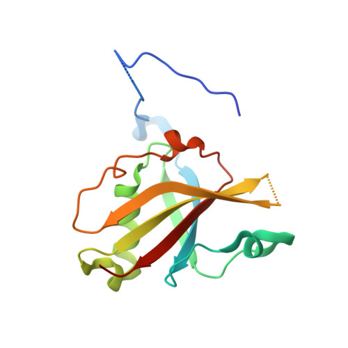
Crystal structure of mouse SAP18 residues 1-143


Experimental Data Snapshot

  • Method: X-RAY DIFFRACTION
  • Resolution: 1.90 Å
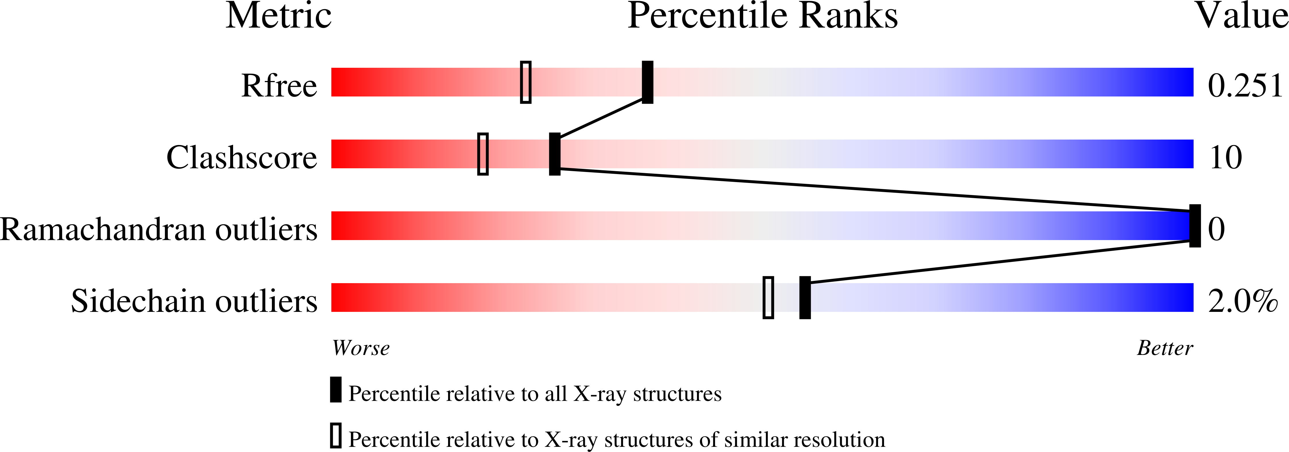
  • R-Value Free: 0.250 
  • R-Value Work: 0.215 
  • R-Value Observed: 0.216 

Starting Model: experimental
View more details

wwPDB Validation   3D Report Full Report


This is version 1.2 of the entry. See complete history


Literature

The Structure of the Asap Core Complex Reveals the Existence of a Pinin-Containing Psap Complex

Murachelli, A.G.Ebert, J.Basquin, C.Le Hir, H.Conti, E.

(2012) Nat Struct Mol Biol 19: 378

  • DOI: https://doi.org/10.1038/nsmb.2242
  • Primary Citation of Related Structures:  
    4A6Q, 4A8X, 4A90

  • PubMed Abstract: 

    The ASAP complex interacts with the exon-junction complex (EJC), a messenger ribonucleoprotein complex involved in post-transcriptional regulation. The three ASAP subunits (Acinus, RNPS1 and SAP18) have been individually implicated in transcriptional regulation, pre-mRNA splicing and mRNA quality control. To shed light on the basis for and consequences of ASAP's interaction with the EJC, we have determined the 1.9-Å resolution structure of a eukaryotic ASAP core complex. The RNA-recognition motif of RNPS1 binds to a conserved motif of Acinus with a recognition mode similar to that observed in splicing U2AF proteins. The Acinus-RNPS1 platform recruits the ubiquitin-like domain of SAP18, forming a ternary complex that has both RNA- and protein-binding properties. Unexpectedly, our structural analysis identified an Acinus-like motif in Pinin, another EJC-associated splicing factor. We show that Pinin physically interacts with RNPS1 and SAP18, forming an alternative ternary complex, PSAP.


  • Organizational Affiliation

    Max Planck Institute of Biochemistry, Department of Structural Cell Biology, Martinsried, Germany.


Macromolecules
Find similar proteins by:  (by identity cutoff)  |  3D Structure
Entity ID: 1
MoleculeChains Sequence LengthOrganismDetailsImage
HISTONE DEACETYLASE COMPLEX SUBUNIT SAP18
A, B
143Mus musculusMutation(s): 0 
UniProt & NIH Common Fund Data Resources
Find proteins for O55128 (Mus musculus)
Explore O55128 
Go to UniProtKB:  O55128
IMPC:  MGI:1277978
Entity Groups  
Sequence Clusters30% Identity50% Identity70% Identity90% Identity95% Identity100% Identity
UniProt GroupO55128
Sequence Annotations
Expand
  • Reference Sequence
Experimental Data & Validation

Experimental Data

  • Method: X-RAY DIFFRACTION
  • Resolution: 1.90 Å
  • R-Value Free: 0.250 
  • R-Value Work: 0.215 
  • R-Value Observed: 0.216 
  • Space Group: C 1 2 1
Unit Cell:
Length ( Å )Angle ( ˚ )
a = 95.42α = 90
b = 65.14β = 119.4
c = 61.61γ = 90
Software Package:
Software NamePurpose
PHENIXrefinement
MOSFLMdata reduction
SCALAdata scaling
PHASERphasing

Structure Validation

View Full Validation Report


Entry History 

Deposition Data

Revision History  (Full details and data files)

  • Version 1.0: 2012-03-07
    Type: Initial release
  • Version 1.1: 2012-04-18
    Changes: Other
  • Version 1.2: 2023-12-20
    Changes: Data collection, Database references, Derived calculations, Other, Refinement description