4YSD

Room temperature structure of copper nitrite reductase from Geobacillus thermodenitrificans


Experimental Data Snapshot

  • Method: X-RAY DIFFRACTION
  • Resolution: 1.35 Å
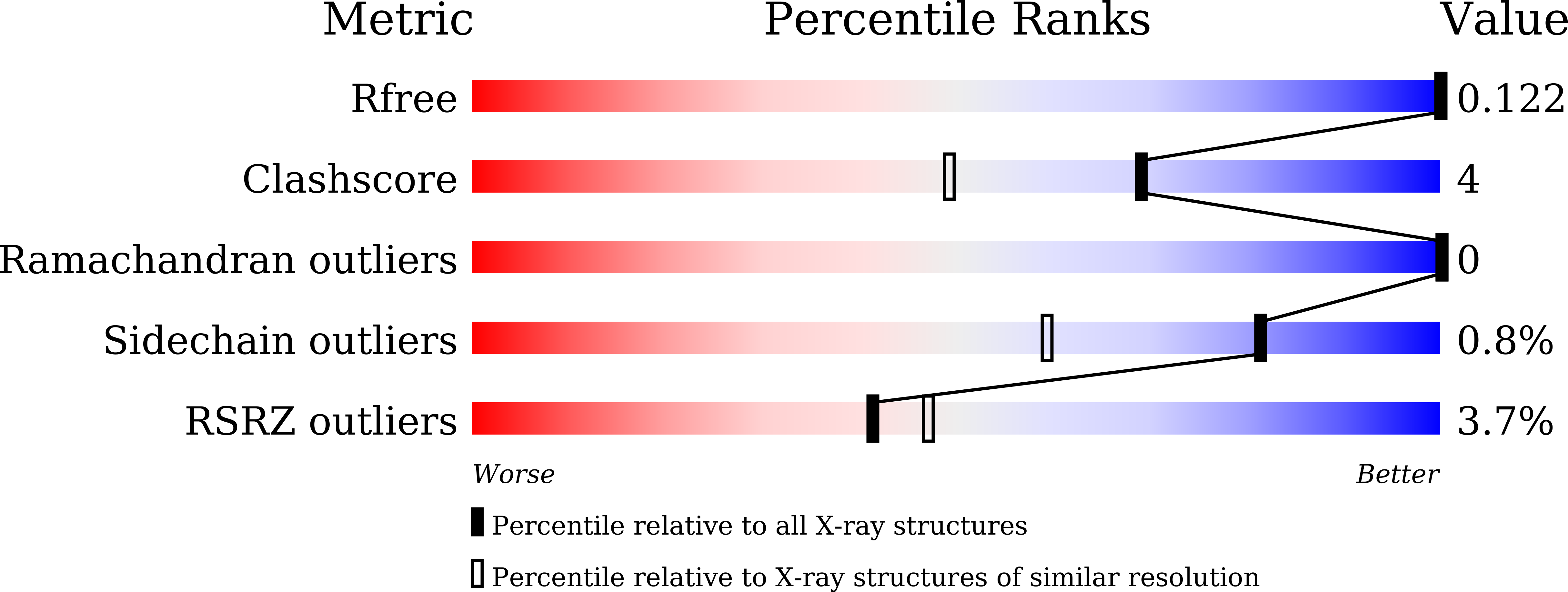
  • R-Value Free: 0.121 
  • R-Value Work: 0.097 
  • R-Value Observed: 0.098 

wwPDB Validation   3D Report Full Report


This is version 1.3 of the entry. See complete history


Literature

Redox-coupled structural changes in nitrite reductase revealed by serial femtosecond and microfocus crystallography

Fukuda, Y.Tse, K.M.Suzuki, M.Diederichs, K.Hirata, K.Nakane, T.Sugahara, M.Nango, E.Tono, K.Joti, Y.Kameshima, T.Song, C.Hatsui, T.Yabashi, M.Nureki, O.Matsumura, H.Inoue, T.Iwata, S.Mizohata, E.

(2016) J Biochem 159: 527-538

  • DOI: https://doi.org/10.1093/jb/mvv133
  • Primary Citation of Related Structures:  
    4YSA, 4YSD, 4YSO, 4YSP, 4YSQ, 4YSR, 4YSS, 4YST, 4YSU

  • PubMed Abstract: 

    Serial femtosecond crystallography (SFX) has enabled the damage-free structural determination of metalloenzymes and filled the gaps of our knowledge between crystallographic and spectroscopic data. Crystallographers, however, scarcely know whether the rising technique provides truly new structural insights into mechanisms of metalloenzymes partly because of limited resolutions. Copper nitrite reductase (CuNiR), which converts nitrite to nitric oxide in denitrification, has been extensively studied by synchrotron radiation crystallography (SRX). Although catalytic Cu (Type 2 copper (T2Cu)) of CuNiR had been suspected to tolerate X-ray photoreduction, we here showed that T2Cu in the form free of nitrite is reduced and changes its coordination structure in SRX. Moreover, we determined the completely oxidized CuNiR structure at 1.43 Å resolution with SFX. Comparison between the high-resolution SFX and SRX data revealed the subtle structural change of a catalytic His residue by X-ray photoreduction. This finding, which SRX has failed to uncover, provides new insight into the reaction mechanism of CuNiR.


  • Organizational Affiliation

    Department of Applied Chemistry, Graduate School of Engineering, Osaka University, 2-1 Yamadaoka, Suita, Osaka 565-0871, Japan;


Macromolecules
Find similar proteins by:  (by identity cutoff)  |  3D Structure
Entity ID: 1
MoleculeChains Sequence LengthOrganismDetailsImage
Nitrite reductase323Geobacillus thermodenitrificans NG80-2Mutation(s): 0 
Gene Names: nirKGTNG_0650
EC: 1.7.2.1
UniProt
Find proteins for A4IL26 (Geobacillus thermodenitrificans (strain NG80-2))
Explore A4IL26 
Go to UniProtKB:  A4IL26
Entity Groups  
Sequence Clusters30% Identity50% Identity70% Identity90% Identity95% Identity100% Identity
UniProt GroupA4IL26
Sequence Annotations
Expand
  • Reference Sequence
Small Molecules
Ligands 2 Unique
IDChains Name / Formula / InChI Key2D Diagram3D Interactions
CU
Query on CU

Download Ideal Coordinates CCD File 
B [auth A]
C [auth A]
D [auth A]
E [auth A]
F [auth A]
B [auth A],
C [auth A],
D [auth A],
E [auth A],
F [auth A],
G [auth A],
H [auth A],
I [auth A],
J [auth A],
K [auth A],
L [auth A],
M [auth A]
COPPER (II) ION
Cu
JPVYNHNXODAKFH-UHFFFAOYSA-N
ACY
Query on ACY

Download Ideal Coordinates CCD File 
N [auth A]ACETIC ACID
C2 H4 O2
QTBSBXVTEAMEQO-UHFFFAOYSA-N
Experimental Data & Validation

Experimental Data

  • Method: X-RAY DIFFRACTION
  • Resolution: 1.35 Å
  • R-Value Free: 0.121 
  • R-Value Work: 0.097 
  • R-Value Observed: 0.098 
  • Space Group: H 3
Unit Cell:
Length ( Å )Angle ( ˚ )
a = 116.197α = 90
b = 116.197β = 90
c = 85.553γ = 120
Software Package:
Software NamePurpose
REFMACrefinement
HKL-2000data reduction
HKL-2000data scaling
MOLREPphasing

Structure Validation

View Full Validation Report


Entry History 

Revision History  (Full details and data files)

  • Version 1.0: 2016-02-24
    Type: Initial release
  • Version 1.1: 2016-05-25
    Changes: Database references
  • Version 1.2: 2020-02-05
    Changes: Data collection, Derived calculations
  • Version 1.3: 2024-03-20
    Changes: Data collection, Database references, Derived calculations