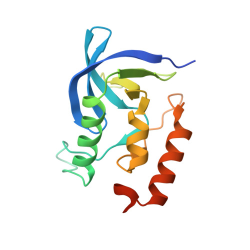
Three-dimensional diffuse x-ray scattering from crystals of Staphylococcal nuclease.
Wall, M.E., Ealick, S.E., Gruner, S.M.(1997) Proc Natl Acad Sci U S A : 6180-6184
- PubMed: 9177191
- DOI: https://doi.org/10.1073/pnas.94.12.6180
- Primary Citation of Related Structures:
4WOR - PubMed Abstract:
We have developed methods for obtaining and characterizing three-dimensional maps of the reciprocal-space distribution of diffuse x-ray scattering from protein crystals, and have used the methods to study the nature of disorder in crystals of Staphylococcal nuclease. Experimentally obtained maps are 99.5% complete in the reciprocal-space resolution range of 10 A-2.5 A, show symmetry consistent with the P41 space group of the unit cell, and are highly reproducible. Quantitative comparisons of the data with three-dimensional simulations imply liquid-like motions of the protein [Caspar, D. L. D., Clarage, J., Salunke, D. M. & Clarage, M. (1988) Nature (London) 332, 659-662], with a correlation length of 10 A and a root-mean-square displacement of 0.36 A.
Organizational Affiliation:
Department of Physics, Princeton University, Princeton, NJ 08544, USA.