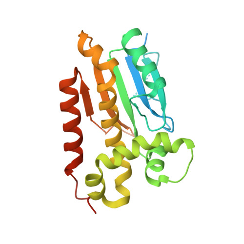
Allosteric Reversion of Haemophilus influenzae beta-Carbonic Anhydrase via a Proline Shift.
Hoffmann, K.M., Million-Perez, H.R., Merkhofer, R., Nicholson, H., Rowlett, R.S.(2015) Biochemistry 54: 598-611
- PubMed: 25506786
- DOI: https://doi.org/10.1021/bi501116e
- Primary Citation of Related Structures:
4WAJ, 4WAK, 4WAM - PubMed Abstract:
Haemophilus influenzae β-carbonic anhydrase (HICA) has been reverse-engineered in the allosteric site region to resemble the nonallosteric Pisum sativum enzyme in order to identify critical features of allostery and intersusbunit communication. Three variants (W39V/G41A, P48S/A49P, and W39V/G41A/P48S/A49P) were identified, through a comparison with a crystal structure of nonallosteric P. sativum β-carbonic anhydrase (PSCA, PDB 1EKJ ), to potentially revert HICA to a nonallosteric enzyme. The W39V/G41A and P48S/A49P mutations decreased the apparent kcat/Km proton dependence from 4 to 2 and 1, respectively, increasing the overall maximal kcat/Km to 16 ± 2 μM(-1) s(-1) (380% of wild type) and 17 ± 3 μM(-1) s(-1) (405% of wild type). The pKa values of the metal-bound water molecule based on the pH-rate profile kinetics (8.32 ± 0.04 for W39V/G41A and 8.3 ± 0.1 for P48S/A49P) were also slightly higher than that for the wild-type enzyme (7.74 ± 0.04). The P48S/A49P variant has lost all pH-rate cooperativity. The W39V/G41A/P48S/A49P variant's kinetics were unusual and were fit with a log-linear function with a slope 0.9 ± 0.2. The crystal structure of the W39V/G41A variant revealed an active site very similar to the T-state wild-type oligomer with bicarbonate trapped in the escort site. By contrast, the X-ray crystal structure of a proline shift variant (P48S/A49P) reveals that it has adopted an active site conformation nearly identical to that of nonallosteric β-carbonic anhydrase (R-state) for one chain, including a tight association with the dimer-exchanged N-terminal helices; the second chain in the asymmetric unit is associated in a biologically relevant oligomer, but it adopts a T-state conformation that is not capped by dimer-exchanged N-terminal helices. The hybrid R/T nature of HICA P48S/A49P structurally recapitulates the interruption of pH-rate cooperativity observed for this variant. Comparison of the conformations of the R and T chains of P48S/A49P suggests a new hypothesis to explain HICA allosteric communication that is mediated by the N-terminal helices and anion binding at the dimer interface.
Organizational Affiliation:
Department of Chemistry, Gonzaga University , 502 East Boone Avenue, Spokane, Washington 99258, United States.