4QD2

Molecular basis for disruption of E-cadherin adhesion by botulinum neurotoxin A complex


Experimental Data Snapshot

  • Method: X-RAY DIFFRACTION
  • Resolution: 2.40 Å
  • R-Value Free: 0.247 
  • R-Value Work: 0.208 
  • R-Value Observed: 0.210 

Starting Models: experimental
View more details

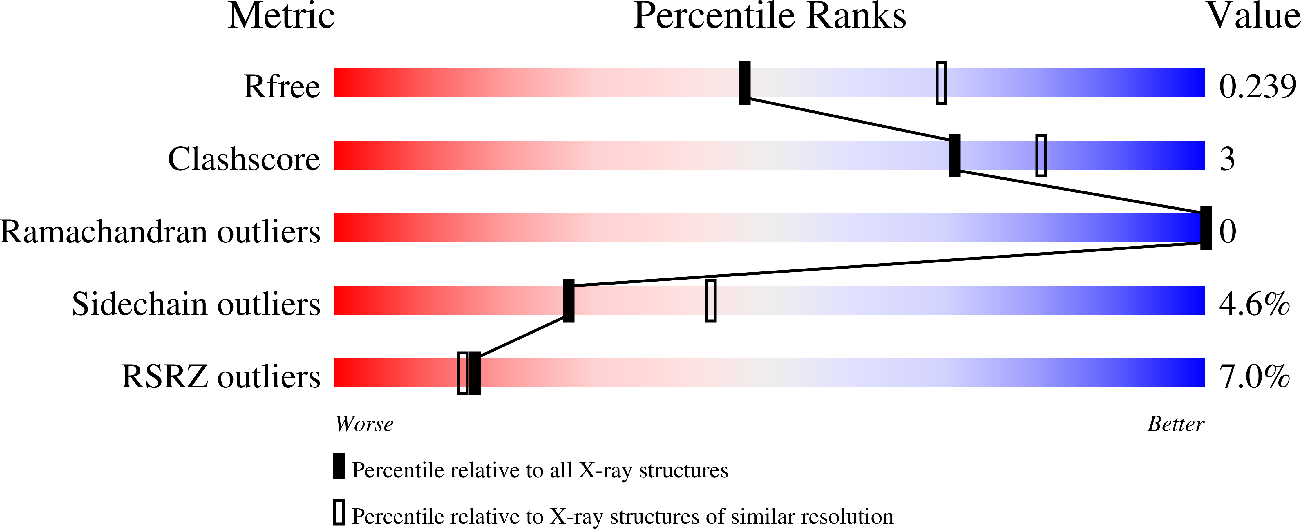
wwPDB Validation   3D Report Full Report


This is version 1.2 of the entry. See complete history


Literature

Molecular basis for disruption of E-cadherin adhesion by botulinum neurotoxin A complex.

Lee, K.Zhong, X.Gu, S.Kruel, A.M.Dorner, M.B.Perry, K.Rummel, A.Dong, M.Jin, R.

(2014) Science 344: 1405-1410

  • DOI: https://doi.org/10.1126/science.1253823
  • Primary Citation of Related Structures:  
    4QD2

  • PubMed Abstract: 

    How botulinum neurotoxins (BoNTs) cross the host intestinal epithelial barrier in foodborne botulism is poorly understood. Here, we present the crystal structure of a clostridial hemagglutinin (HA) complex of serotype BoNT/A bound to the cell adhesion protein E-cadherin at 2.4 angstroms. The HA complex recognizes E-cadherin with high specificity involving extensive intermolecular interactions and also binds to carbohydrates on the cell surface. Binding of the HA complex sequesters E-cadherin in the monomeric state, compromising the E-cadherin-mediated intercellular barrier and facilitating paracellular absorption of BoNT/A. We reconstituted the complete 14-subunit BoNT/A complex using recombinantly produced components and demonstrated that abolishing either E-cadherin- or carbohydrate-binding of the HA complex drastically reduces oral toxicity of BoNT/A complex in vivo. Together, these studies establish the molecular mechanism of how HAs contribute to the oral toxicity of BoNT/A.


  • Organizational Affiliation

    Department of Physiology and Biophysics, University of California, Irvine, CA 92697, USA.


Macromolecules
Find similar proteins by:  (by identity cutoff)  |  3D Structure
Entity ID: 1
MoleculeChains Sequence LengthOrganismDetailsImage
Hemagglutinin component HA33A [auth D],
C,
F [auth I],
H
296Clostridium botulinum A str. HallMutation(s): 0 
Gene Names: ha70CBO0801CLC_0857
UniProt
Find proteins for A5HZZ6 (Clostridium botulinum (strain Hall / ATCC 3502 / NCTC 13319 / Type A))
Explore A5HZZ6 
Go to UniProtKB:  A5HZZ6
Entity Groups  
Sequence Clusters30% Identity50% Identity70% Identity90% Identity95% Identity100% Identity
UniProt GroupA5HZZ6
Sequence Annotations
Expand
  • Reference Sequence
Find similar proteins by:  (by identity cutoff)  |  3D Structure
Entity ID: 2
MoleculeChains Sequence LengthOrganismDetailsImage
Hemagglutinin component HA17
B, G
147Clostridium botulinum A str. HallMutation(s): 0 
Gene Names: ha17CBO0802CLC_0858
UniProt
Find proteins for A5HZZ5 (Clostridium botulinum (strain Hall / ATCC 3502 / NCTC 13319 / Type A))
Explore A5HZZ5 
Go to UniProtKB:  A5HZZ5
Entity Groups  
Sequence Clusters30% Identity50% Identity70% Identity90% Identity95% Identity100% Identity
UniProt GroupA5HZZ5
Sequence Annotations
Expand
  • Reference Sequence
Find similar proteins by:  (by identity cutoff)  |  3D Structure
Entity ID: 3
MoleculeChains Sequence LengthOrganismDetailsImage
Hemagglutinin component HA70D [auth A],
I [auth F]
254Clostridium botulinum A str. HallMutation(s): 0 
Gene Names: ha33CBO0803CLC_0859
UniProt
Find proteins for A5HZZ4 (Clostridium botulinum (strain Hall / ATCC 3502 / NCTC 13319 / Type A))
Explore A5HZZ4 
Go to UniProtKB:  A5HZZ4
Entity Groups  
Sequence Clusters30% Identity50% Identity70% Identity90% Identity95% Identity100% Identity
UniProt GroupA5HZZ4
Sequence Annotations
Expand
  • Reference Sequence
Find similar proteins by:  (by identity cutoff)  |  3D Structure
Entity ID: 4
MoleculeChains Sequence LengthOrganismDetailsImage
Cadherin-1
E, J
213Mus musculusMutation(s): 0 
Gene Names: Cdh1
UniProt & NIH Common Fund Data Resources
Find proteins for P09803 (Mus musculus)
Explore P09803 
Go to UniProtKB:  P09803
IMPC:  MGI:88354
Entity Groups  
Sequence Clusters30% Identity50% Identity70% Identity90% Identity95% Identity100% Identity
UniProt GroupP09803
Sequence Annotations
Expand
  • Reference Sequence
Experimental Data & Validation

Experimental Data

  • Method: X-RAY DIFFRACTION
  • Resolution: 2.40 Å
  • R-Value Free: 0.247 
  • R-Value Work: 0.208 
  • R-Value Observed: 0.210 
  • Space Group: P 1
Unit Cell:
Length ( Å )Angle ( ˚ )
a = 86.99α = 116.89
b = 95.73β = 96.34
c = 96.14γ = 93.7
Software Package:
Software NamePurpose
ADSCdata collection
PHASERphasing
PHENIXrefinement
MOSFLMdata reduction
SCALAdata scaling

Structure Validation

View Full Validation Report


Entry History 

Deposition Data

Revision History  (Full details and data files)

  • Version 1.0: 2014-06-25
    Type: Initial release
  • Version 1.1: 2014-07-02
    Changes: Database references
  • Version 1.2: 2023-09-20
    Changes: Data collection, Database references, Derived calculations, Refinement description