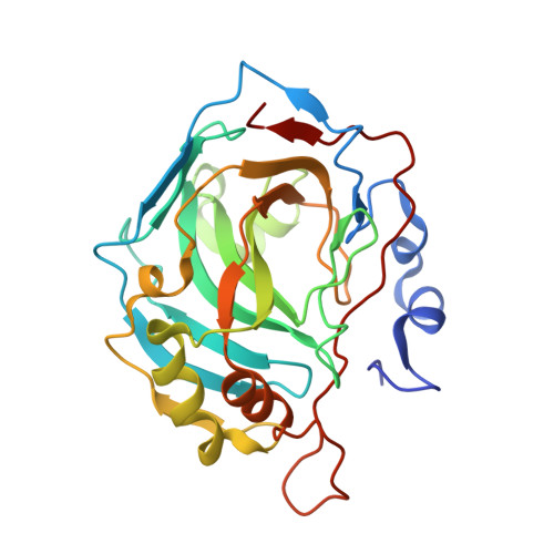
Exploring the influence of the protein environment on metal-binding pharmacophores.
Martin, D.P., Blachly, P.G., McCammon, J.A., Cohen, S.M.(2014) J Med Chem 57: 7126-7135
- PubMed: 25116076
- DOI: https://doi.org/10.1021/jm500984b
- Primary Citation of Related Structures:
4Q7P, 4Q7S, 4Q7V, 4Q7W, 4Q81, 4Q83, 4Q87, 4Q8X, 4Q8Y, 4Q8Z, 4Q90, 4Q99, 4Q9Y - PubMed Abstract:
The binding of a series of metal-binding pharmacophores (MBPs) related to the ligand 1-hydroxypyridine-2-(1H)-thione (1,2-HOPTO) in the active site of human carbonic anhydrase II (hCAII) has been investigated. The presence and/or position of a single methyl substituent drastically alters inhibitor potency and can result in coordination modes not observed in small-molecule model complexes. It is shown that this unexpected binding mode is the result of a steric clash between the methyl group and a highly ordered water network in the active site that is further stabilized by the formation of a hydrogen bond and favorable hydrophobic contacts. The affinity of MBPs is dependent on a large number of factors including donor atom identity, orientation, electrostatics, and van der Waals interactions. These results suggest that metal coordination by metalloenzyme inhibitors is a malleable interaction and that it is thus more appropriate to consider the metal-binding motif of these inhibitors as a pharmacophore rather than a "chelator". The rational design of inhibitors targeting metalloenzymes will benefit greatly from a deeper understanding of the interplay between the variety of forces governing the binding of MBPs to active site metal ions.
Organizational Affiliation:
Departments of Chemistry and Biochemistry, ‡Pharmacology, and §Howard Hughes Medical Institute, University of California, San Diego , 9500 Gilman Drive, MC 0358, La Jolla, California 92093, United States.


