4PEI

Dbr1 in complex with synthetic branched RNA analog


Experimental Data Snapshot

  • Method: X-RAY DIFFRACTION
  • Resolution: 1.95 Å
  • R-Value Free: 0.229 
  • R-Value Work: 0.184 
  • R-Value Observed: 0.186 

wwPDB Validation   3D Report Full Report


This is version 1.5 of the entry. See complete history


Literature

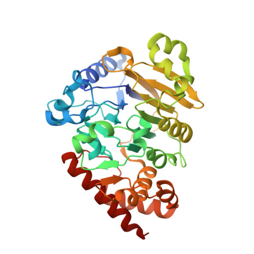
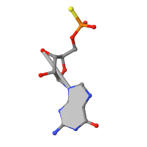
Structural basis of lariat RNA recognition by the intron debranching enzyme Dbr1.

Montemayor, E.J.Katolik, A.Clark, N.E.Taylor, A.B.Schuermann, J.P.Combs, D.J.Johnsson, R.Holloway, S.P.Stevens, S.W.Damha, M.J.Hart, P.J.

(2014) Nucleic Acids Res 42: 10845-10855

  • DOI: https://doi.org/10.1093/nar/gku725
  • Primary Citation of Related Structures:  
    4PEF, 4PEG, 4PEH, 4PEI

  • PubMed Abstract: 

    The enzymatic processing of cellular RNA molecules requires selective recognition of unique chemical and topological features. The unusual 2',5'-phosphodiester linkages in RNA lariats produced by the spliceosome must be hydrolyzed by the intron debranching enzyme (Dbr1) before they can be metabolized or processed into essential cellular factors, such as snoRNA and miRNA. Dbr1 is also involved in the propagation of retrotransposons and retroviruses, although the precise role played by the enzyme in these processes is poorly understood. Here, we report the first structures of Dbr1 alone and in complex with several synthetic RNA compounds that mimic the branchpoint in lariat RNA. The structures, together with functional data on Dbr1 variants, reveal the molecular basis for 2',5'-phosphodiester recognition and explain why the enzyme lacks activity toward 3',5'-phosphodiester linkages. The findings illuminate structure/function relationships in a unique enzyme that is central to eukaryotic RNA metabolism and set the stage for the rational design of inhibitors that may represent novel therapeutic agents to treat retroviral infections and neurodegenerative disease.


  • Organizational Affiliation

    Department of Biochemistry, The University of Texas Health Science Center, San Antonio, TX 78229, USA X-ray Crystallography Core Laboratory, The University of Texas Health Science Center, San Antonio, TX 78229, USA.


Macromolecules

Find similar proteins by:  (by identity cutoff)  |  3D Structure
Entity ID: 1
MoleculeChains Sequence LengthOrganismDetailsImage
RNA lariat debranching enzyme, putativeA,
D [auth B],
G [auth C],
J [auth D],
M [auth E]
356Entamoeba histolyticaMutation(s): 1 
Gene Names: EHI_062730
UniProt
Find proteins for C4M1P9 (Entamoeba histolytica (strain ATCC 30459 / HM-1:IMSS / ABRM))
Explore C4M1P9 
Go to UniProtKB:  C4M1P9
Entity Groups  
Sequence Clusters30% Identity50% Identity70% Identity90% Identity95% Identity100% Identity
UniProt GroupC4M1P9
Sequence Annotations
Expand
  • Reference Sequence

Find similar nucleic acids by:  Sequence   |   3D Structure  

Entity ID: 2
MoleculeChains LengthOrganismImage
RNA (5'-R(*UP*AP*AP*CP*A)-3')B [auth V],
E [auth W],
H [auth X],
K [auth Y],
N [auth Z]
5synthetic construct
Sequence Annotations
Expand
  • Reference Sequence

Find similar nucleic acids by:  Sequence   |   3D Structure  

Entity ID: 3
MoleculeChains LengthOrganismImage
RNA (5'-R(*(G46)P*U)-3')C [auth Q],
F [auth R],
I [auth S],
L [auth T],
O [auth U]
2synthetic construct
Sequence Annotations
Expand
  • Reference Sequence
Small Molecules
Ligands 4 Unique
IDChains Name / Formula / InChI Key2D Diagram3D Interactions
PG4
Query on PG4

Download Ideal Coordinates CCD File 
V [auth B]TETRAETHYLENE GLYCOL
C8 H18 O5
UWHCKJMYHZGTIT-UHFFFAOYSA-N
SO4
Query on SO4

Download Ideal Coordinates CCD File 
BA [auth D],
FA [auth E],
Q [auth A],
U [auth B],
X [auth C]
SULFATE ION
O4 S
QAOWNCQODCNURD-UHFFFAOYSA-L
GOL
Query on GOL

Download Ideal Coordinates CCD File 
CA [auth D]
DA [auth D]
R [auth A]
S [auth A]
Y [auth C]
CA [auth D],
DA [auth D],
R [auth A],
S [auth A],
Y [auth C],
Z [auth C]
GLYCEROL
C3 H8 O3
PEDCQBHIVMGVHV-UHFFFAOYSA-N
NI
Query on NI

Download Ideal Coordinates CCD File 
AA [auth D],
EA [auth E],
P [auth A],
T [auth B],
W [auth C]
NICKEL (II) ION
Ni
VEQPNABPJHWNSG-UHFFFAOYSA-N
Experimental Data & Validation

Experimental Data

  • Method: X-RAY DIFFRACTION
  • Resolution: 1.95 Å
  • R-Value Free: 0.229 
  • R-Value Work: 0.184 
  • R-Value Observed: 0.186 
  • Space Group: P 21 21 21
Unit Cell:
Length ( Å )Angle ( ˚ )
a = 73.037α = 90
b = 142.479β = 90
c = 214.665γ = 90
Software Package:
Software NamePurpose
PHENIXrefinement

Structure Validation

View Full Validation Report


Entry History 

Revision History  (Full details and data files)

  • Version 1.0: 2014-08-27
    Type: Initial release
  • Version 1.1: 2014-10-01
    Changes: Database references
  • Version 1.2: 2014-11-12
    Changes: Atomic model, Derived calculations
  • Version 1.3: 2015-02-25
    Changes: Non-polymer description
  • Version 1.4: 2017-09-27
    Changes: Advisory, Author supporting evidence, Derived calculations, Other, Source and taxonomy, Structure summary
  • Version 1.5: 2023-12-27
    Changes: Data collection, Database references, Derived calculations