4ORN

Blue Fluorescent Protein mKalama1


Experimental Data Snapshot

  • Method: X-RAY DIFFRACTION
  • Resolution: 1.71 Å
  • R-Value Free: 0.183 
  • R-Value Work: 0.158 
  • R-Value Observed: 0.159 

wwPDB Validation   3D Report Full Report


This is version 2.0 of the entry. See complete history


Literature

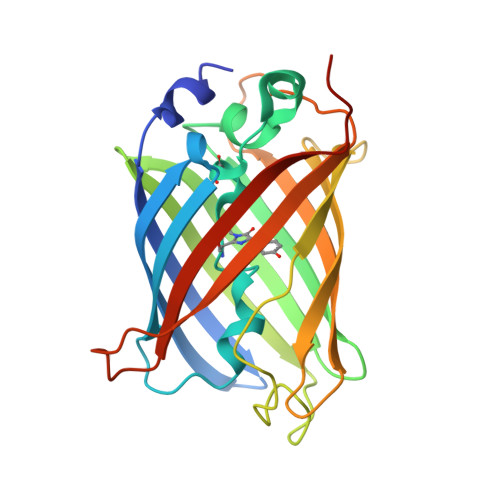
Hidden photoinduced reactivity of the blue fluorescent protein mKalama1.

Vegh, R.B.Bloch, D.A.Bommarius, A.S.Verkhovsky, M.Pletnev, S.Iwai, H.Bochenkova, A.V.Solntsev, K.M.

(2015) Phys Chem Chem Phys 17: 12472-12485

  • DOI: https://doi.org/10.1039/c5cp00887e
  • Primary Citation of Related Structures:  
    4ORN

  • PubMed Abstract: 

    Understanding the photoinduced dynamics of fluorescent proteins is essential for their applications in bioimaging. Despite numerous studies on the ultrafast dynamics, the delayed response of these proteins, which often results in population of kinetically trapped dark states of various origins, is largely unexplored. Here, by using transient absorption spectroscopy spanning the time scale from picoseconds to seconds, we reveal a hidden reactivity of the bright blue-light emitting protein mKalama1 previously thought to be inert. This protein shows no excited-state proton transfer during its nanosecond excited-state lifetime; however, its tyrosine-based chromophore undergoes deprotonation coupled to non-radiative electronic relaxation. Such deprotonation causes distinct optical absorption changes in the broad UV-to-NIR spectral range (ca. 300-800 nm); the disappearance of the transient absorption signal has a complex nature and spans the whole microsecond-to-second time scale. The mechanisms underlying the relaxation kinetics are disclosed based on the X-ray structural analysis of mKalama1 and the high-level electronic structure calculations of proposed intermediates in the photocycle. We conclude that the non-radiative excited-state decay includes two major branches: internal conversion coupled to intraprotein proton transfer, where a conserved residue E222 serves as the proton acceptor; and ionization induced by two consecutive resonant absorption events, followed by deprotonation of the chromophore radical cation to bulk solvent through a novel water-mediated proton-wire pathway. Our findings open up new perspectives on the dynamics of fluorescent proteins as tracked by its optical transient absorption in the time domain extending up to seconds.


  • Organizational Affiliation

    School of Chemistry and Biochemistry, Georgia Institute of Technology, Atlanta, Georgia 30332, USA. solntsev@gatech.edu.


Macromolecules
Find similar proteins by:  (by identity cutoff)  |  3D Structure
Entity ID: 1
MoleculeChains Sequence LengthOrganismDetailsImage
mKalama1
A, B
239Aequorea victoriaMutation(s): 25 
Gene Names: GFP
UniProt
Find proteins for P42212 (Aequorea victoria)
Explore P42212 
Go to UniProtKB:  P42212
Entity Groups  
Sequence Clusters30% Identity50% Identity70% Identity90% Identity95% Identity100% Identity
UniProt GroupP42212
Sequence Annotations
Expand
  • Reference Sequence
Small Molecules
Ligands 3 Unique
IDChains Name / Formula / InChI Key2D Diagram3D Interactions
MES
Query on MES

Download Ideal Coordinates CCD File 
C [auth A],
D [auth A]
2-(N-MORPHOLINO)-ETHANESULFONIC ACID
C6 H13 N O4 S
SXGZJKUKBWWHRA-UHFFFAOYSA-N
SO4
Query on SO4

Download Ideal Coordinates CCD File 
E [auth A]
F [auth A]
G [auth A]
I [auth B]
J [auth B]
E [auth A],
F [auth A],
G [auth A],
I [auth B],
J [auth B],
K [auth B]
SULFATE ION
O4 S
QAOWNCQODCNURD-UHFFFAOYSA-L
CL
Query on CL

Download Ideal Coordinates CCD File 
H [auth A],
L [auth B],
M [auth B],
N [auth B]
CHLORIDE ION
Cl
VEXZGXHMUGYJMC-UHFFFAOYSA-M
Modified Residues  2 Unique
IDChains TypeFormula2D DiagramParent
CSD
Query on CSD
A, B
L-PEPTIDE LINKINGC3 H7 N O4 SCYS
GYS
Query on GYS
A, B
L-PEPTIDE LINKINGC14 H15 N3 O5SER, TYR, GLY
Experimental Data & Validation

Experimental Data

  • Method: X-RAY DIFFRACTION
  • Resolution: 1.71 Å
  • R-Value Free: 0.183 
  • R-Value Work: 0.158 
  • R-Value Observed: 0.159 
  • Space Group: P 21 21 21
Unit Cell:
Length ( Å )Angle ( ˚ )
a = 80.51α = 90
b = 83.46β = 90
c = 92.24γ = 90
Software Package:
Software NamePurpose
XSCALEdata scaling
PHENIXrefinement
PDB_EXTRACTdata extraction
MOSFLMdata reduction
PHASERphasing

Structure Validation

View Full Validation Report


Entry History 

Deposition Data

Revision History  (Full details and data files)

  • Version 1.0: 2015-02-11
    Type: Initial release
  • Version 1.1: 2015-04-08
    Changes: Database references
  • Version 1.2: 2015-05-20
    Changes: Database references
  • Version 1.3: 2023-09-20
    Changes: Data collection, Database references, Derived calculations, Refinement description
  • Version 2.0: 2023-11-15
    Changes: Atomic model, Data collection, Derived calculations