


Crystal structure of putative glycogen phosphorylase from Streptococcus mutans
Lihan, M.-Y., Li, G.-L., Li, L.-F., Su, X.-D.To be published.
Experimental Data Snapshot
Starting Model: experimental
View more details
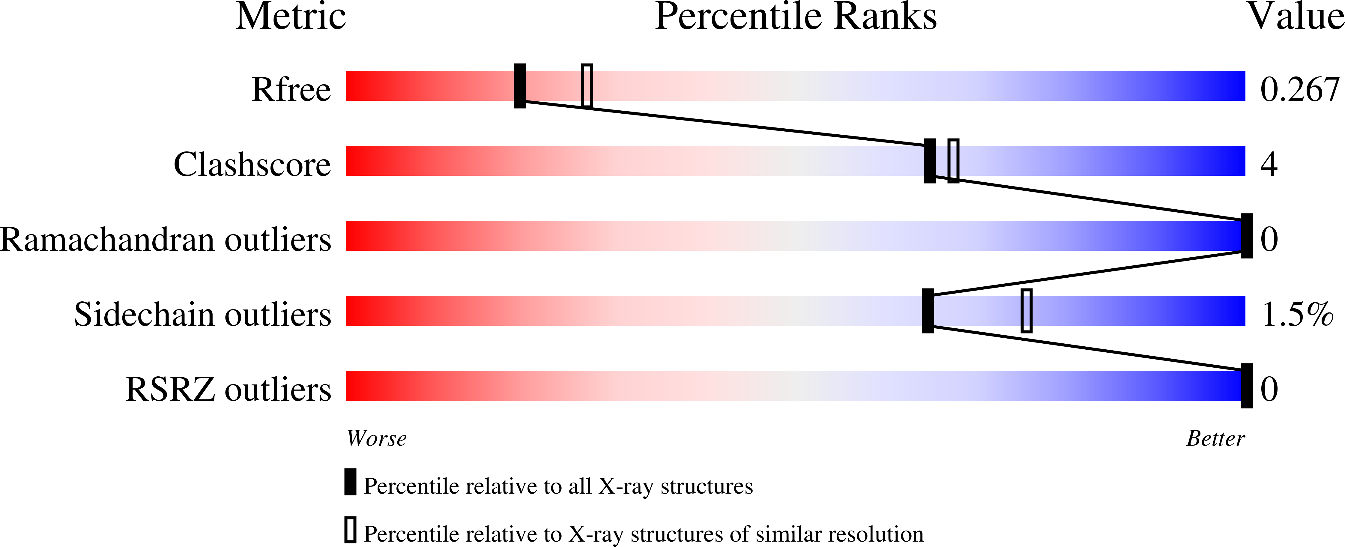
wwPDB Validation 3D Report Full Report
Entity ID: 1 | |||||
|---|---|---|---|---|---|
| Molecule | Chains | Sequence Length | Organism | Details | Image |
| Phosphorylase | 758 | Streptococcus mutans UA159 | Mutation(s): 0 Gene Names: glgP, SMU_1564 EC: 2.4.1.1 | 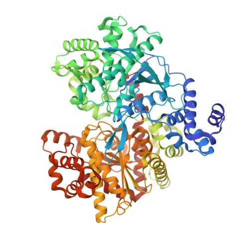 | |
UniProt | |||||
Find proteins for Q8DT31 (Streptococcus mutans serotype c (strain ATCC 700610 / UA159)) Explore Q8DT31 Go to UniProtKB: Q8DT31 | |||||
Entity Groups | |||||
| Sequence Clusters | 30% Identity50% Identity70% Identity90% Identity95% Identity100% Identity | ||||
| UniProt Group | Q8DT31 | ||||
Sequence AnnotationsExpand | |||||
| |||||
| Length ( Å ) | Angle ( ˚ ) |
|---|---|
| a = 119.72 | α = 90 |
| b = 85.02 | β = 93.3 |
| c = 84.46 | γ = 90 |
| Software Name | Purpose |
|---|---|
| REFMAC | refinement |
| PHENIX | refinement |
| HKL-2000 | data reduction |
| HKL-2000 | data scaling |
| HKL-2000 | data collection |
RCSB PDB (citation) is hosted by
RCSB PDB is a member of the

