4JVU

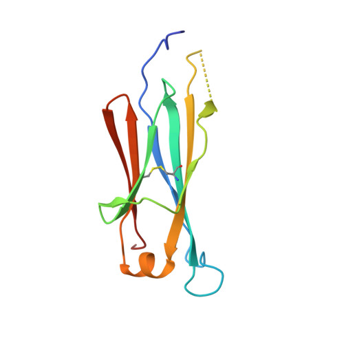
IgM C2-domain from mouse


Experimental Data Snapshot

  • Method: X-RAY DIFFRACTION
  • Resolution: 1.30 Å
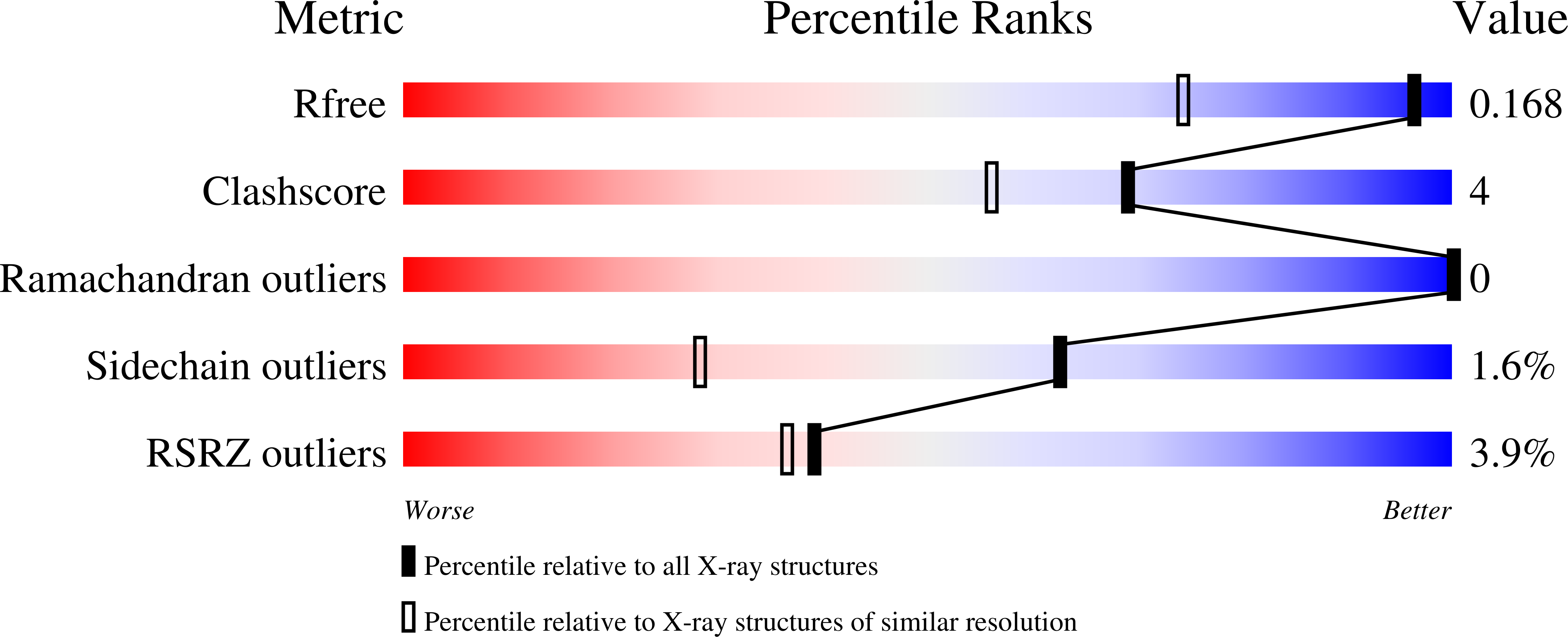
  • R-Value Free: 0.168 
  • R-Value Work: 0.138 
  • R-Value Observed: 0.139 

Starting Model: experimental
View more details

wwPDB Validation   3D Report Full Report


This is version 1.3 of the entry. See complete history


Literature

High-resolution structures of the IgM Fc domains reveal principles of its hexamer formation.

Muller, R.Grawert, M.A.Kern, T.Madl, T.Peschek, J.Sattler, M.Groll, M.Buchner, J.

(2013) Proc Natl Acad Sci U S A 110: 10183-10188

  • DOI: https://doi.org/10.1073/pnas.1300547110
  • Primary Citation of Related Structures:  
    4BA8, 4JVU, 4JVW

  • PubMed Abstract: 

    IgM is the first antibody produced during the humoral immune response. Despite its fundamental role in the immune system, IgM is structurally only poorly described. In this work we used X-ray crystallography and NMR spectroscopy to determine the atomic structures of the constant IgM Fc domains (Cµ2, Cµ3, and Cµ4) and to address their roles in IgM oligomerization. Although the isolated domains share the typical Ig fold, they differ substantially in dimerization properties and quaternary contacts. Unexpectedly, the Cµ4 domain and its C-terminal tail piece are responsible and sufficient for the specific polymerization of Cµ4 dimers into covalently linked hexamers of dimers. Based on small angle X-ray scattering data, we present a model of the ring-shaped Cµ4 structure, which reveals the principles of IgM oligomerization.


  • Organizational Affiliation

    Munich Center for Integrated Protein Science at the Department Chemie, Technische Universität München, 85748 Garching, Germany.


Macromolecules
Find similar proteins by:  (by identity cutoff)  |  3D Structure
Entity ID: 1
MoleculeChains Sequence LengthOrganismDetailsImage
Ig mu chain C region membrane-bound form
A, B
117Mus musculusMutation(s): 0 
Gene Names: Igh-6
UniProt
Find proteins for P01872 (Mus musculus)
Explore P01872 
Go to UniProtKB:  P01872
Entity Groups  
Sequence Clusters30% Identity50% Identity70% Identity90% Identity95% Identity100% Identity
UniProt GroupP01872
Sequence Annotations
Expand
  • Reference Sequence
Experimental Data & Validation

Experimental Data

  • Method: X-RAY DIFFRACTION
  • Resolution: 1.30 Å
  • R-Value Free: 0.168 
  • R-Value Work: 0.138 
  • R-Value Observed: 0.139 
  • Space Group: C 1 2 1
Unit Cell:
Length ( Å )Angle ( ˚ )
a = 92.44α = 90
b = 44.78β = 118.1
c = 54.57γ = 90
Software Package:
Software NamePurpose
REFMACrefinement
XDSdata scaling
XSCALEdata scaling
PHASERphasing

Structure Validation

View Full Validation Report


Entry History 

Deposition Data

Revision History  (Full details and data files)

  • Version 1.0: 2013-06-12
    Type: Initial release
  • Version 1.1: 2013-07-03
    Changes: Database references
  • Version 1.2: 2023-09-20
    Changes: Data collection, Database references, Refinement description
  • Version 1.3: 2024-10-30
    Changes: Structure summary