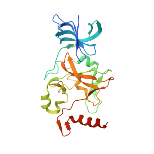
4JLG

SETD7 in complex with inhibitor (R)-PFI-2 and S-adenosyl-methionine


Experimental Data Snapshot

  • Method: X-RAY DIFFRACTION
  • Resolution: 1.90 Å
  • R-Value Free: 0.237 
  • R-Value Work: 0.211 
  • R-Value Observed: 0.212 

Starting Model: experimental
View more details

wwPDB Validation   3D Report Full Report


Ligand Structure Quality Assessment 


This is version 1.3 of the entry. See complete history


Literature

(R)-PFI-2 is a potent and selective inhibitor of SETD7 methyltransferase activity in cells.

Barsyte-Lovejoy, D.Li, F.Oudhoff, M.J.Tatlock, J.H.Dong, A.Zeng, H.Wu, H.Freeman, S.A.Schapira, M.Senisterra, G.A.Kuznetsova, E.Marcellus, R.Allali-Hassani, A.Kennedy, S.Lambert, J.P.Couzens, A.L.Aman, A.Gingras, A.C.Al-Awar, R.Fish, P.V.Gerstenberger, B.S.Roberts, L.Benn, C.L.Grimley, R.L.Braam, M.J.Rossi, F.M.Sudol, M.Brown, P.J.Bunnage, M.E.Owen, D.R.Zaph, C.Vedadi, M.Arrowsmith, C.H.

(2014) Proc Natl Acad Sci U S A 111: 12853-12858

  • DOI: https://doi.org/10.1073/pnas.1407358111
  • Primary Citation of Related Structures:  
    4JLG

  • PubMed Abstract: 

    SET domain containing (lysine methyltransferase) 7 (SETD7) is implicated in multiple signaling and disease related pathways with a broad diversity of reported substrates. Here, we report the discovery of (R)-PFI-2-a first-in-class, potent (Ki (app) = 0.33 nM), selective, and cell-active inhibitor of the methyltransferase activity of human SETD7-and its 500-fold less active enantiomer, (S)-PFI-2. (R)-PFI-2 exhibits an unusual cofactor-dependent and substrate-competitive inhibitory mechanism by occupying the substrate peptide binding groove of SETD7, including the catalytic lysine-binding channel, and by making direct contact with the donor methyl group of the cofactor, S-adenosylmethionine. Chemoproteomics experiments using a biotinylated derivative of (R)-PFI-2 demonstrated dose-dependent competition for binding to endogenous SETD7 in MCF7 cells pretreated with (R)-PFI-2. In murine embryonic fibroblasts, (R)-PFI-2 treatment phenocopied the effects of Setd7 deficiency on Hippo pathway signaling, via modulation of the transcriptional coactivator Yes-associated protein (YAP) and regulation of YAP target genes. In confluent MCF7 cells, (R)-PFI-2 rapidly altered YAP localization, suggesting continuous and dynamic regulation of YAP by the methyltransferase activity of SETD7. These data establish (R)-PFI-2 and related compounds as a valuable tool-kit for the study of the diverse roles of SETD7 in cells and further validate protein methyltransferases as a druggable target class.


  • Organizational Affiliation

    Structural Genomics Consortium, University of Toronto, Toronto, ON, Canada M5G 1L7; carrow@uhnres.utoronto.ca m.vedadi@utoronto.ca d.barsyte@utoronto.ca.


Macromolecules
Find similar proteins by:  (by identity cutoff)  |  3D Structure
Entity ID: 1
MoleculeChains Sequence LengthOrganismDetailsImage
Histone-lysine N-methyltransferase SETD7
A, B
264Homo sapiensMutation(s): 0 
Gene Names: SETD7KIAA1717KMT7SET7SET9
EC: 2.1.1.43 (PDB Primary Data), 2.1.1.364 (UniProt)
UniProt & NIH Common Fund Data Resources
Find proteins for Q8WTS6 (Homo sapiens)
Explore Q8WTS6 
Go to UniProtKB:  Q8WTS6
PHAROS:  Q8WTS6
GTEx:  ENSG00000145391 
Entity Groups  
Sequence Clusters30% Identity50% Identity70% Identity90% Identity95% Identity100% Identity
UniProt GroupQ8WTS6
Sequence Annotations
Expand
  • Reference Sequence
Small Molecules
Ligands 3 Unique
IDChains Name / Formula / InChI Key2D Diagram3D Interactions
1L8
Query on 1L8

Download Ideal Coordinates CCD File 
D [auth A],
W [auth B]
8-fluoro-N-{(2R)-1-oxo-1-(pyrrolidin-1-yl)-3-[3-(trifluoromethyl)phenyl]propan-2-yl}-1,2,3,4-tetrahydroisoquinoline-6-sulfonamide
C23 H25 F4 N3 O3 S
JCKGSPAAPQRPBW-OAQYLSRUSA-N
SAM
Query on SAM

Download Ideal Coordinates CCD File 
C [auth A],
V [auth B]
S-ADENOSYLMETHIONINE
C15 H22 N6 O5 S
MEFKEPWMEQBLKI-FCKMPRQPSA-N
UNX
Query on UNX

Download Ideal Coordinates CCD File 
AA [auth B]
BA [auth B]
CA [auth B]
DA [auth B]
E [auth A]
AA [auth B],
BA [auth B],
CA [auth B],
DA [auth B],
E [auth A],
EA [auth B],
F [auth A],
G [auth A],
H [auth A],
I [auth A],
J [auth A],
K [auth A],
L [auth A],
M [auth A],
N [auth A],
O [auth A],
P [auth A],
Q [auth A],
R [auth A],
S [auth A],
T [auth A],
U [auth A],
X [auth B],
Y [auth B],
Z [auth B]
UNKNOWN ATOM OR ION
X
Binding Affinity Annotations 
IDSourceBinding Affinity
1L8 BindingDB:  4JLG Ki: 0.33 (nM) from 1 assay(s)
IC50: 2 (nM) from 1 assay(s)
Experimental Data & Validation

Experimental Data

  • Method: X-RAY DIFFRACTION
  • Resolution: 1.90 Å
  • R-Value Free: 0.237 
  • R-Value Work: 0.211 
  • R-Value Observed: 0.212 
  • Space Group: P 1
Unit Cell:
Length ( Å )Angle ( ˚ )
a = 46.742α = 83.33
b = 57.984β = 71.42
c = 64.932γ = 70.93
Software Package:
Software NamePurpose
SCALEPACKdata scaling
REFMACrefinement
PDB_EXTRACTdata extraction
JBluIce-EPICSdata collection
HKL-2000data reduction
PHASESphasing

Structure Validation

View Full Validation Report


Ligand Structure Quality Assessment 


Entry History 

Revision History  (Full details and data files)

  • Version 1.0: 2013-04-17
    Type: Initial release
  • Version 1.1: 2014-04-09
    Changes: Structure summary
  • Version 1.2: 2015-02-18
    Changes: Database references
  • Version 1.3: 2023-09-20
    Changes: Data collection, Database references, Derived calculations, Refinement description