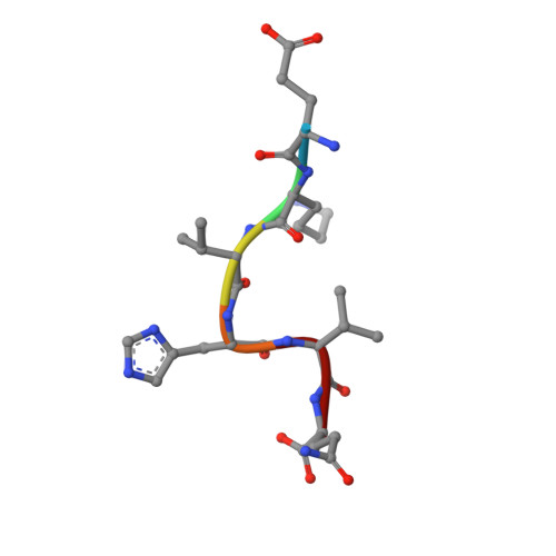
4J79

Crystal structure of beta'-COP/PEDVspike complex


Experimental Data Snapshot

  • Method: X-RAY DIFFRACTION
  • Resolution: 1.56 Å
  • R-Value Free: 0.196 
  • R-Value Work: 0.165 
  • R-Value Observed: 0.166 

wwPDB Validation   3D Report Full Report


This is version 1.2 of the entry. See complete history


Literature

Rules for the recognition of dilysine retrieval motifs by coatomer.

Ma, W.Goldberg, J.

(2013) EMBO J 32: 926-937

  • DOI: https://doi.org/10.1038/emboj.2013.41
  • Primary Citation of Related Structures:  
    4J73, 4J77, 4J78, 4J79, 4J81, 4J82, 4J84, 4J86, 4J87, 4J8B, 4J8G

  • PubMed Abstract: 

    Cytoplasmic dilysine motifs on transmembrane proteins are captured by coatomer α-COP and β'-COP subunits and packaged into COPI-coated vesicles for Golgi-to-ER retrieval. Numerous ER/Golgi proteins contain K(x)Kxx motifs, but the rules for their recognition are unclear. We present crystal structures of α-COP and β'-COP bound to a series of naturally occurring retrieval motifs-encompassing KKxx, KxKxx and non-canonical RKxx and viral KxHxx sequences. Binding experiments show that α-COP and β'-COP have generally the same specificity for KKxx and KxKxx, but only β'-COP recognizes the RKxx signal. Dilysine motif recognition involves lysine side-chain interactions with two acidic patches. Surprisingly, however, KKxx and KxKxx motifs bind differently, with their lysine residues transposed at the binding patches. We derive rules for retrieval motif recognition from key structural features: the reversed binding modes, the recognition of the C-terminal carboxylate group which enforces lysine positional context, and the tolerance of the acidic patches for non-lysine residues.


  • Organizational Affiliation

    Memorial Sloan-Kettering Cancer Center, Howard Hughes Medical Institute and the Structural Biology Program, New York, NY, USA.


Macromolecules
Find similar proteins by:  (by identity cutoff)  |  3D Structure
Entity ID: 1
MoleculeChains Sequence LengthOrganismDetailsImage
Coatomer subunit beta'301Saccharomyces cerevisiaeMutation(s): 0 
UniProt
Find proteins for P41811 (Saccharomyces cerevisiae (strain ATCC 204508 / S288c))
Explore P41811 
Go to UniProtKB:  P41811
Entity Groups  
Sequence Clusters30% Identity50% Identity70% Identity90% Identity95% Identity100% Identity
UniProt GroupP41811
Sequence Annotations
Expand
  • Reference Sequence

Find similar proteins by:  Sequence   |   3D Structure  

Entity ID: 2
MoleculeChains Sequence LengthOrganismDetailsImage
Spike glycoprotein6Porcine epidemic diarrhea virusMutation(s): 0 
UniProt
Find proteins for Q91AV1 (Porcine epidemic diarrhea virus (strain CV777))
Explore Q91AV1 
Go to UniProtKB:  Q91AV1
Entity Groups  
UniProt GroupQ91AV1
Sequence Annotations
Expand
  • Reference Sequence
Experimental Data & Validation

Experimental Data

  • Method: X-RAY DIFFRACTION
  • Resolution: 1.56 Å
  • R-Value Free: 0.196 
  • R-Value Work: 0.165 
  • R-Value Observed: 0.166 
  • Space Group: C 1 2 1
Unit Cell:
Length ( Å )Angle ( ˚ )
a = 75.1α = 90
b = 51.18β = 101.75
c = 85.25γ = 90
Software Package:
Software NamePurpose
PHENIXrefinement

Structure Validation

View Full Validation Report


Entry History 

Deposition Data

Revision History  (Full details and data files)

  • Version 1.0: 2013-03-27
    Type: Initial release
  • Version 1.1: 2013-04-17
    Changes: Database references
  • Version 1.2: 2024-02-28
    Changes: Data collection, Database references