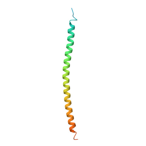
4I1L

Structural and Biological Features of FOXP3 Dimerization Relevant to Regulatory T Cell Function


Experimental Data Snapshot

  • Method: X-RAY DIFFRACTION
  • Resolution: 2.10 Å
  • R-Value Free: 0.289 
  • R-Value Work: 0.236 
  • R-Value Observed: 0.238 

wwPDB Validation   3D Report Full Report


This is version 1.1 of the entry. See complete history


Literature

Structural and biological features of FOXP3 dimerization relevant to regulatory T cell function.

Song, X.Li, B.Xiao, Y.Chen, C.Wang, Q.Liu, Y.Berezov, A.Xu, C.Gao, Y.Li, Z.Wu, S.L.Cai, Z.Zhang, H.Karger, B.L.Hancock, W.W.Wells, A.D.Zhou, Z.Greene, M.I.

(2012) Cell Rep 1: 665-675

  • DOI: https://doi.org/10.1016/j.celrep.2012.04.012
  • Primary Citation of Related Structures:  
    4I1L

  • PubMed Abstract: 

    FOXP3 is a key transcription factor for regulatory T cell function. We report the crystal structure of the FOXP3 coiled-coil domain, through which a loose or transient dimeric association is formed and modulated, accounting for the activity variations introduced by disease-causing mutations or posttranslational modifications. Structure-guided mutagenesis revealed that FOXP3 coiled-coil-mediated homodimerization is essential for Treg function in vitro and in vivo. In particular, we identified human FOXP3 K250 and K252 as key residues for the conformational change and stability of the FOXP3 dimer, which can be regulated by protein posttranslational modifications such as reversible lysine acetylation. These studies provide structural and mechanistic explanations for certain disease-causing mutations in the coiled-coil domain of FOXP3 that are commonly found in IPEX syndrome. Overall, the regulatory machinery involving homooligomerization, acetylation, and heteroassociation has been dissected, defining atomic insights into the biological and pathological characteristics of the FOXP3 complex.


  • Organizational Affiliation

    State Key Laboratory of Cell Biology, Institute of Biochemistry and Cell Biology, Chinese Academy of Sciences, Shanghai, China 200031.


Macromolecules
Find similar proteins by:  (by identity cutoff)  |  3D Structure
Entity ID: 1
MoleculeChains Sequence LengthOrganismDetailsImage
Forkhead box protein P393Mus musculusMutation(s): 0 
Gene Names: Foxp3
UniProt
Find proteins for Q99JB6 (Mus musculus)
Explore Q99JB6 
Go to UniProtKB:  Q99JB6
Entity Groups  
Sequence Clusters30% Identity50% Identity70% Identity90% Identity95% Identity100% Identity
UniProt GroupQ99JB6
Sequence Annotations
Expand
  • Reference Sequence
Small Molecules
Ligands 3 Unique
IDChains Name / Formula / InChI Key2D Diagram3D Interactions
SO4
Query on SO4

Download Ideal Coordinates CCD File 
B [auth A],
C [auth A],
D [auth A]
SULFATE ION
O4 S
QAOWNCQODCNURD-UHFFFAOYSA-L
ACT
Query on ACT

Download Ideal Coordinates CCD File 
H [auth A],
I [auth A]
ACETATE ION
C2 H3 O2
QTBSBXVTEAMEQO-UHFFFAOYSA-M
MG
Query on MG

Download Ideal Coordinates CCD File 
E [auth A],
F [auth A],
G [auth A]
MAGNESIUM ION
Mg
JLVVSXFLKOJNIY-UHFFFAOYSA-N
Experimental Data & Validation

Experimental Data

  • Method: X-RAY DIFFRACTION
  • Resolution: 2.10 Å
  • R-Value Free: 0.289 
  • R-Value Work: 0.236 
  • R-Value Observed: 0.238 
  • Space Group: P 43 2 2
Unit Cell:
Length ( Å )Angle ( ˚ )
a = 37.13α = 90
b = 37.13β = 90
c = 135.672γ = 90
Software Package:
Software NamePurpose
ADSCdata collection
SOLVEphasing
REFMACrefinement
HKL-2000data reduction
HKL-2000data scaling

Structure Validation

View Full Validation Report


Entry History 

Deposition Data

Revision History  (Full details and data files)

  • Version 1.0: 2012-12-05
    Type: Initial release
  • Version 1.1: 2024-03-20
    Changes: Data collection, Database references, Derived calculations