4EW9

The liganded structure of C. bescii family 3 pectate lyase


Experimental Data Snapshot

  • Method: X-RAY DIFFRACTION
  • Resolution: 1.60 Å
  • R-Value Free: 0.204 
  • R-Value Work: 0.164 
  • R-Value Observed: 0.166 

wwPDB Validation   3D Report Full Report


Ligand Structure Quality Assessment 


This is version 3.1 of the entry. See complete history


Literature

The structure and mode of action of Caldicellulosiruptor bescii family 3 pectate lyase in biomass deconstruction.

Alahuhta, M.Brunecky, R.Chandrayan, P.Kataeva, I.Adams, M.W.Himmel, M.E.Lunin, V.V.

(2013) Acta Crystallogr D Biol Crystallogr 69: 534-539

  • DOI: https://doi.org/10.1107/S0907444912050512
  • Primary Citation of Related Structures:  
    4EW9

  • PubMed Abstract: 

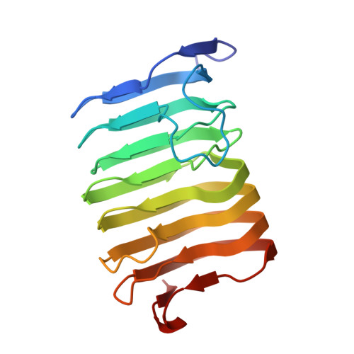
    The unique active site of the Caldicellulosiruptor bescii family 3 pectate lyase catalytic module (PL3-cat) has been structurally described and synergistic digestion studies with C. bescii cellulase A have been performed on unpretreated biomass. The X-ray structure of PL3-cat was determined at 1.6 Å resolution (PDB entry 4ew9) in complex with the products of trigalacturonic acid. Comparison with family 1 pectate lyase (PL1) structures shows that the active site of the PL3 catalytic module is considerably different. However, on superimposing the identical sugar rings at the -2 subsites conserved interactions could be identified. Interestingly, only one catalytic residue, the lysine that donates the proton to the carboxylate group in the β-elimination reaction of PL1 (Lys108 in PL3-cat), is conserved in PL3 and there is no arginine to abstract the proton from the C5 carbon of the galactouronate ring. This suggests that the reaction mechanism of PL3 requires different catalytic residues. Most interestingly, comparison with other proton-abstraction reactions reveals that in PL3 the α-proton is abstracted by a lysine, in a striking similarity to enolases. These observations led us to propose that in PL3-cat Lys108 is the catalytic base, Glu84 is the catalytic acid and an acidified water molecule completes the anti β-elimination reaction by protonating the O4 atom of the substrate. Also, our digestion experiments with unpretreated switchgrass show that the loadings of C. bescii cellobiohydrolase A (CelA) can be lowered by the addition of PL3 to the reaction mixture. This result suggests that PL3 can significantly improve the deconstruction of unpretreated biomass by allowing other enzymes to better access their preferred substrates.


  • Organizational Affiliation

    Biosciences Center, National Renewable Energy Laboratory, Golden, CO 80401-3305, USA.


Macromolecules
Find similar proteins by:  (by identity cutoff)  |  3D Structure
Entity ID: 1
MoleculeChains Sequence LengthOrganismDetailsImage
Pectate lyase
A, B
195Caldicellulosiruptor bescii DSM 6725Mutation(s): 0 
Gene Names: Athe_1854
EC: 4.2.2.2
UniProt
Find proteins for B9MKT4 (Caldicellulosiruptor bescii (strain ATCC BAA-1888 / DSM 6725 / KCTC 15123 / Z-1320))
Explore B9MKT4 
Go to UniProtKB:  B9MKT4
Entity Groups  
Sequence Clusters30% Identity50% Identity70% Identity90% Identity95% Identity100% Identity
UniProt GroupB9MKT4
Sequence Annotations
Expand
  • Reference Sequence
Oligosaccharides

Help

Entity ID: 2
MoleculeChains Length2D Diagram Glycosylation3D Interactions
4-deoxy-beta-L-threo-hex-4-enopyranuronic acid-(1-4)-alpha-D-galactopyranuronic acid
C, D
2N/A
Glycosylation Resources
GlyTouCan:  G31388FK
GlyCosmos:  G31388FK
Small Molecules
Ligands 5 Unique
IDChains Name / Formula / InChI Key2D Diagram3D Interactions
ADA
Query on ADA

Download Ideal Coordinates CCD File 
N [auth A]alpha-D-galactopyranuronic acid
C6 H10 O7
AEMOLEFTQBMNLQ-BKBMJHBISA-N
DGU
Query on DGU

Download Ideal Coordinates CCD File 
M [auth A],
U [auth B]
D-galacturonic acid
C6 H10 O7
IAJILQKETJEXLJ-RSJOWCBRSA-N
MPD
Query on MPD

Download Ideal Coordinates CCD File 
J [auth A],
K [auth A],
L [auth A],
T [auth B]
(4S)-2-METHYL-2,4-PENTANEDIOL
C6 H14 O2
SVTBMSDMJJWYQN-YFKPBYRVSA-N
ACT
Query on ACT

Download Ideal Coordinates CCD File 
O [auth A],
V [auth B]
ACETATE ION
C2 H3 O2
QTBSBXVTEAMEQO-UHFFFAOYSA-M
CA
Query on CA

Download Ideal Coordinates CCD File 
E [auth A]
F [auth A]
G [auth A]
H [auth A]
I [auth A]
E [auth A],
F [auth A],
G [auth A],
H [auth A],
I [auth A],
P [auth B],
Q [auth B],
R [auth B],
S [auth B]
CALCIUM ION
Ca
BHPQYMZQTOCNFJ-UHFFFAOYSA-N
Experimental Data & Validation

Experimental Data

  • Method: X-RAY DIFFRACTION
  • Resolution: 1.60 Å
  • R-Value Free: 0.204 
  • R-Value Work: 0.164 
  • R-Value Observed: 0.166 
  • Space Group: C 1 2 1
Unit Cell:
Length ( Å )Angle ( ˚ )
a = 136.946α = 90
b = 35.898β = 132
c = 98.687γ = 90
Software Package:
Software NamePurpose
PROTEUM PLUSdata collection
PHASERphasing
REFMACrefinement
PROTEUM PLUSdata reduction
PROTEUM PLUSdata scaling

Structure Validation

View Full Validation Report


Ligand Structure Quality Assessment 


Entry History 

Deposition Data

Revision History  (Full details and data files)

  • Version 1.0: 2013-03-20
    Type: Initial release
  • Version 1.1: 2019-06-05
    Changes: Data collection, Database references
  • Version 2.0: 2020-07-01
    Changes: Atomic model, Data collection, Database references, Derived calculations
  • Version 3.0: 2020-07-29
    Type: Remediation
    Reason: Carbohydrate remediation
    Changes: Advisory, Atomic model, Data collection, Derived calculations, Non-polymer description, Structure summary
  • Version 3.1: 2023-09-13
    Changes: Data collection, Database references, Refinement description, Structure summary