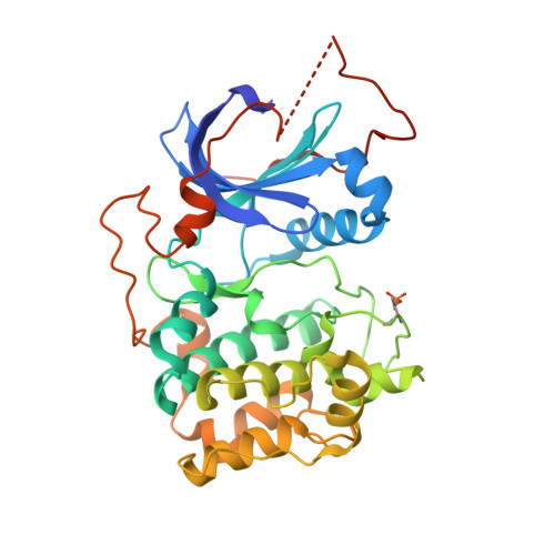
4EKL

Akt1 with GDC0068


Experimental Data Snapshot

  • Method: X-RAY DIFFRACTION
  • Resolution: 2.00 Å
  • R-Value Free: 0.235 
  • R-Value Work: 0.172 
  • R-Value Observed: 0.175 

wwPDB Validation   3D Report Full Report


Ligand Structure Quality Assessment 


This is version 1.1 of the entry. See complete history


Literature

An ATP-Site On-Off Switch That Restricts Phosphatase Accessibility of Akt.

Lin, K.Lin, J.Wu, W.I.Ballard, J.Lee, B.B.Gloor, S.L.Vigers, G.P.Morales, T.H.Friedman, L.S.Skelton, N.Brandhuber, B.J.

(2012) Sci Signal 5: ra37-ra37

  • DOI: https://doi.org/10.1126/scisignal.2002618
  • Primary Citation of Related Structures:  
    4EKK, 4EKL

  • PubMed Abstract: 

    The protein serine-threonine kinase Akt undergoes a substantial conformational change upon activation, which is induced by the phosphorylation of two critical regulatory residues, threonine 308 and serine 473. Paradoxically, treating cells with adenosine 5'-triphosphate (ATP)-competitive inhibitors of Akt results in increased phosphorylation of both residues. We show that binding of ATP-competitive inhibitors stabilized a conformation in which both phosphorylated sites were inaccessible to phosphatases. ATP binding also produced this protection of the phosphorylated sites, whereas interaction with its hydrolysis product adenosine 5'-diphosphate (ADP) or allosteric Akt inhibitors resulted in increased accessibility of these phosphorylated residues. ATP-competitive inhibitors mimicked ATP by targeting active Akt. Forms of Akt activated by an oncogenic mutation or myristoylation were more potently inhibited by the ATP-competitive inhibitors than was wild-type Akt. These data support a new model of kinase regulation, wherein nucleotides modulate an on-off switch in Akt through conformational changes, which is disrupted by ATP-competitive inhibitors.


  • Organizational Affiliation

    Genentech Inc., South San Francisco, CA 94080, USA. klin@gene.com


Macromolecules
Find similar proteins by:  (by identity cutoff)  |  3D Structure
Entity ID: 1
MoleculeChains Sequence LengthOrganismDetailsImage
RAC-alpha serine/threonine-protein kinase341Homo sapiensMutation(s): 1 
Gene Names: AKT1PKBRAC
EC: 2.7.11.1
UniProt & NIH Common Fund Data Resources
Find proteins for P31749 (Homo sapiens)
Explore P31749 
Go to UniProtKB:  P31749
PHAROS:  P31749
GTEx:  ENSG00000142208 
Entity Groups  
Sequence Clusters30% Identity50% Identity70% Identity90% Identity95% Identity100% Identity
UniProt GroupP31749
Sequence Annotations
Expand
  • Reference Sequence
Small Molecules
Ligands 1 Unique
IDChains Name / Formula / InChI Key2D Diagram3D Interactions
0RF
Query on 0RF

Download Ideal Coordinates CCD File 
B [auth A](2S)-2-(4-chlorophenyl)-1-{4-[(5R,7R)-7-hydroxy-5-methyl-6,7-dihydro-5H-cyclopenta[d]pyrimidin-4-yl]piperazin-1-yl}-3-(propan-2-ylamino)propan-1-one
C24 H32 Cl N5 O2
GRZXWCHAXNAUHY-NSISKUIASA-N
Modified Residues  1 Unique
IDChains TypeFormula2D DiagramParent
TPO
Query on TPO
A
L-PEPTIDE LINKINGC4 H10 N O6 PTHR
Binding Affinity Annotations 
IDSourceBinding Affinity
0RF BindingDB:  4EKL Kd: min: 0.64, max: 35 (nM) from 3 assay(s)
IC50: min: 1.3, max: 157 (nM) from 16 assay(s)
Experimental Data & Validation

Experimental Data

  • Method: X-RAY DIFFRACTION
  • Resolution: 2.00 Å
  • R-Value Free: 0.235 
  • R-Value Work: 0.172 
  • R-Value Observed: 0.175 
  • Space Group: P 21 21 21
Unit Cell:
Length ( Å )Angle ( ˚ )
a = 47.031α = 90
b = 57.949β = 90
c = 151.61γ = 90
Software Package:
Software NamePurpose
CrystalCleardata collection
MOLREPphasing
REFMACrefinement
MOSFLMdata reduction
SCALAdata scaling

Structure Validation

View Full Validation Report


Ligand Structure Quality Assessment 


Entry History 

Deposition Data

Revision History  (Full details and data files)

  • Version 1.0: 2012-05-23
    Type: Initial release
  • Version 1.1: 2018-01-24
    Changes: Structure summary