Two Amino Acid Residues Confer Different Binding Affinities of Abelson Family Kinase Src Homology 2 Domains for Phosphorylated Cortactin.
Gifford, S.M., Liu, W., Mader, C.C., Halo, T.L., Machida, K., Boggon, T.J., Koleske, A.J.(2014) J Biol Chem 289: 19704-19713
- PubMed: 24891505
- DOI: https://doi.org/10.1074/jbc.M114.556480
- Primary Citation of Related Structures:
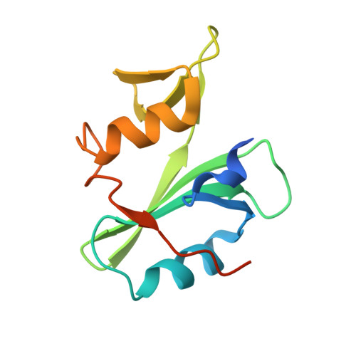
4EIH - PubMed Abstract:
The closely related Abl family kinases, Arg and Abl, play important non-redundant roles in the regulation of cell morphogenesis and motility. Despite similar N-terminal sequences, Arg and Abl interact with different substrates and binding partners with varying affinities. This selectivity may be due to slight differences in amino acid sequence leading to differential interactions with target proteins. We report that the Arg Src homology (SH) 2 domain binds two specific phosphotyrosines on cortactin, a known Abl/Arg substrate, with over 10-fold higher affinity than the Abl SH2 domain. We show that this significant affinity difference is due to the substitution of arginine 161 and serine 187 in Abl to leucine 207 and threonine 233 in Arg, respectively. We constructed Abl SH2 domains with R161L and S187T mutations alone and in combination and find that these substitutions are sufficient to convert the low affinity Abl SH2 domain to a higher affinity "Arg-like" SH2 domain in binding to a phospho-cortactin peptide. We crystallized the Arg SH2 domain for structural comparison to existing crystal structures of the Abl SH2 domain. We show that these two residues are important determinants of Arg and Abl SH2 domain binding specificity. Finally, we expressed Arg containing an "Abl-like" low affinity mutant Arg SH2 domain (L207R/T233S) and find that this mutant, although properly localized to the cell periphery, does not support wild type levels of cell edge protrusion. Together, these observations indicate that these two amino acid positions confer different binding affinities and cellular functions on the distinct Abl family kinases.
Organizational Affiliation:
From the Departments of Molecular Biophysics and Biochemistry.