4CCY

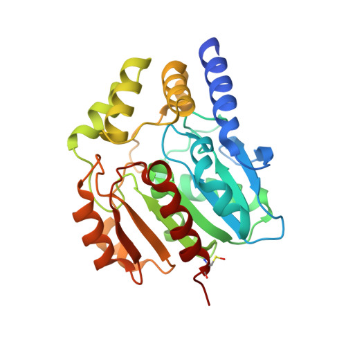
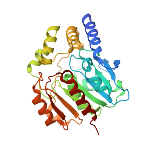
Crystal structure of carboxylesterase CesB (YbfK) from Bacillus subtilis


Experimental Data Snapshot

  • Method: X-RAY DIFFRACTION
  • Resolution: 2.04 Å
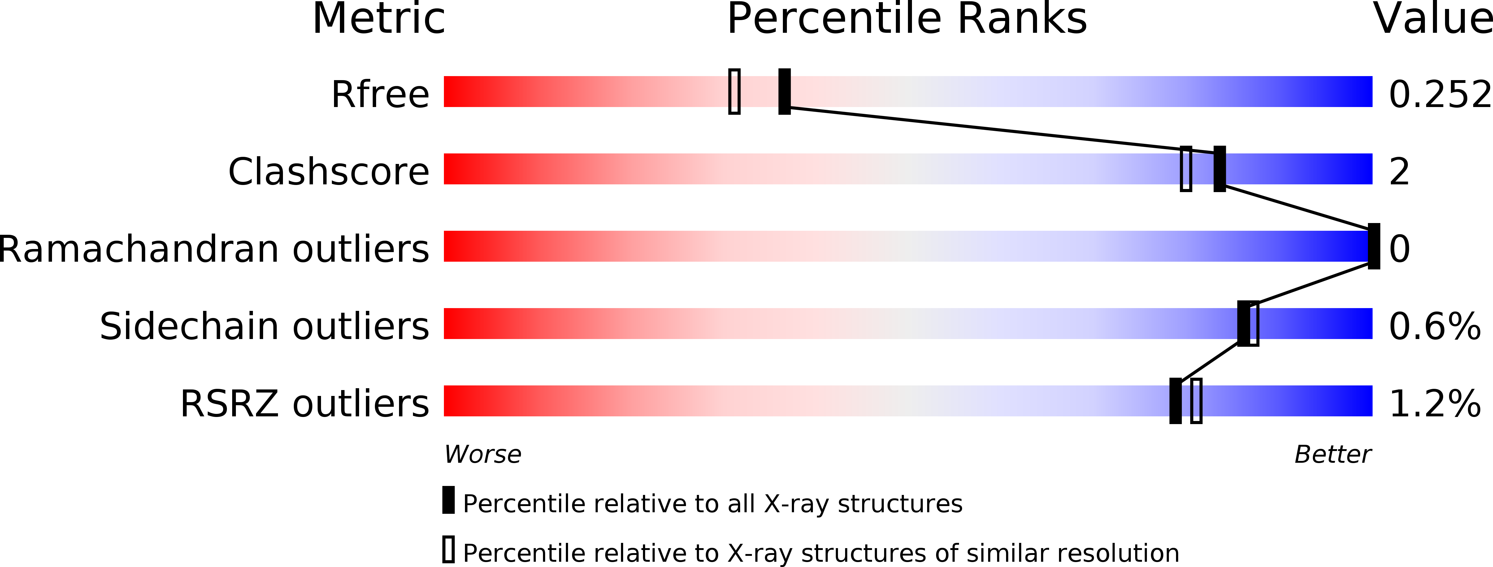
  • R-Value Free: 0.250 
  • R-Value Work: 0.204 
  • R-Value Observed: 0.206 

Starting Model: experimental
View more details

wwPDB Validation   3D Report Full Report


This is version 1.5 of the entry. See complete history


Literature

Crystal Structures of Two Bacillus Carboxylesterases with Different Enantioselectivities.

Rozeboom, H.J.Godinho, L.F.Nardini, M.Quax, W.J.Dijkstra, B.W.

(2014) Biochim Biophys Acta 1844: 567

  • DOI: https://doi.org/10.1016/j.bbapap.2014.01.003
  • Primary Citation of Related Structures:  
    4CCW, 4CCY

  • PubMed Abstract: 

    Naproxen esterase (NP) from Bacillus subtilis Thai I-8 is a carboxylesterase that catalyzes the enantioselective hydrolysis of naproxenmethylester to produce S-naproxen (E>200). It is a homolog of CesA (98% sequence identity) and CesB (64% identity), both produced by B. subtilis strain 168. CesB can be used for the enantioselective hydrolysis of 1,2-O-isopropylideneglycerol (solketal) esters (E>200 for IPG-caprylate). Crystal structures of NP and CesB, determined to a resolution of 1.75Å and 2.04Å, respectively, showed that both proteins have a canonical α/β hydrolase fold with an extra N-terminal helix stabilizing the cap subdomain. The active site in both enzymes is located in a deep hydrophobic groove and includes the catalytic triad residues Ser130, His274, and Glu245. A product analog, presumably 2-(2-hydroxyethoxy)acetic acid, was bound in the NP active site. The enzymes have different enantioselectivities, which previously were shown to result from only a few amino acid substitutions in the cap domain. Modeling of a substrate in the active site of NP allowed explaining the different enantioselectivities. In addition, Ala156 may be a determinant of enantioselectivity as well, since its side chain appears to interfere with the binding of certain R-enantiomers in the active site of NP. However, the exchange route for substrate and product between the active site and the solvent is not obvious from the structures. Flexibility of the cap domain might facilitate such exchange. Interestingly, both carboxylesterases show higher structural similarity to meta-cleavage compound (MCP) hydrolases than to other α/β hydrolase fold esterases.


  • Organizational Affiliation

    Laboratory of Biophysical Chemistry, Centre of Life Sciences, University of Groningen, Nijenborgh 7, 9747 AG Groningen, The Netherlands.


Macromolecules
Find similar proteins by:  (by identity cutoff)  |  3D Structure
Entity ID: 1
MoleculeChains Sequence LengthOrganismDetailsImage
CARBOXYLESTERASE YBFK296Bacillus subtilis subsp. subtilis str. 168Mutation(s): 0 
EC: 3.1.1.1
UniProt
Find proteins for O31452 (Bacillus subtilis (strain 168))
Explore O31452 
Go to UniProtKB:  O31452
Entity Groups  
Sequence Clusters30% Identity50% Identity70% Identity90% Identity95% Identity100% Identity
UniProt GroupO31452
Sequence Annotations
Expand
  • Reference Sequence
Find similar proteins by:  (by identity cutoff)  |  3D Structure
Entity ID: 2
MoleculeChains Sequence LengthOrganismDetailsImage
CARBOXYLESTERASE YBFK296Bacillus subtilis subsp. subtilis str. 168Mutation(s): 0 
EC: 3.1.1.1
UniProt
Find proteins for O31452 (Bacillus subtilis (strain 168))
Explore O31452 
Go to UniProtKB:  O31452
Entity Groups  
Sequence Clusters30% Identity50% Identity70% Identity90% Identity95% Identity100% Identity
UniProt GroupO31452
Sequence Annotations
Expand
  • Reference Sequence
Experimental Data & Validation

Experimental Data

  • Method: X-RAY DIFFRACTION
  • Resolution: 2.04 Å
  • R-Value Free: 0.250 
  • R-Value Work: 0.204 
  • R-Value Observed: 0.206 
  • Space Group: P 1 2 1
Unit Cell:
Length ( Å )Angle ( ˚ )
a = 82.11α = 90
b = 44.01β = 91.87
c = 108.66γ = 90
Software Package:
Software NamePurpose
REFMACrefinement
iMOSFLMdata reduction
SCALAdata scaling
PHASERphasing

Structure Validation

View Full Validation Report


Entry History 

Deposition Data

Revision History  (Full details and data files)

  • Version 1.0: 2014-01-22
    Type: Initial release
  • Version 1.1: 2014-08-13
    Changes: Database references
  • Version 1.2: 2017-07-05
    Changes: Data collection
  • Version 1.3: 2019-05-08
    Changes: Data collection, Derived calculations, Experimental preparation
  • Version 1.4: 2023-12-20
    Changes: Data collection, Database references, Derived calculations, Other, Refinement description
  • Version 1.5: 2024-10-16
    Changes: Structure summary