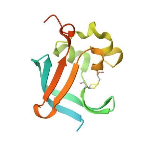
4B0E

Crystal structure of the Caf1A usher protein N-terminal domain from Yersinia pestis


Experimental Data Snapshot

  • Method: X-RAY DIFFRACTION
  • Resolution: 2.00 Å
  • R-Value Free: 0.261 
  • R-Value Work: 0.205 
  • R-Value Observed: 0.205 

Starting Model: other
View more details

wwPDB Validation   3D Report Full Report


This is version 1.5 of the entry. See complete history


Literature

Allosteric Mechanism Controls Traffic in the Chaperone/Usher Pathway.

Di Yu, X.Dubnovitsky, A.Pudney, A.F.Macintyre, S.Knight, S.D.Zavialov, A.V.

(2012) Structure 20: 1861

  • DOI: https://doi.org/10.1016/j.str.2012.08.016
  • Primary Citation of Related Structures:  
    4AY0, 4AYF, 4AZ8, 4B0E, 4B0M

  • PubMed Abstract: 

    Many virulence organelles of Gram-negative bacterial pathogens are assembled via the chaperone/usher pathway. The chaperone transports organelle subunits across the periplasm to the outer membrane usher, where they are released and incorporated into growing fibers. Here, we elucidate the mechanism of the usher-targeting step in assembly of the Yersinia pestis F1 capsule at the atomic level. The usher interacts almost exclusively with the chaperone in the chaperone:subunit complex. In free chaperone, a pair of conserved proline residues at the beginning of the subunit-binding loop form a "proline lock" that occludes the usher-binding surface and blocks usher binding. Binding of the subunit to the chaperone rotates the proline lock away from the usher-binding surface, allowing the chaperone-subunit complex to bind to the usher. We show that the proline lock exists in other chaperone/usher systems and represents a general allosteric mechanism for selective targeting of chaperone:subunit complexes to the usher and for release and recycling of the free chaperone.


  • Organizational Affiliation

    Department of Molecular Biology, Uppsala BioCenter, Swedish University of Agricultural Sciences, BMC, Box 590, SE-75324 Uppsala, Sweden.


Macromolecules
Find similar proteins by:  (by identity cutoff)  |  3D Structure
Entity ID: 1
MoleculeChains Sequence LengthOrganismDetailsImage
F1 CAPSULE-ANCHORING PROTEIN
A, B, C, D
136Yersinia pestisMutation(s): 0 
UniProt
Find proteins for P26949 (Yersinia pestis)
Explore P26949 
Go to UniProtKB:  P26949
Entity Groups  
Sequence Clusters30% Identity50% Identity70% Identity90% Identity95% Identity100% Identity
UniProt GroupP26949
Sequence Annotations
Expand
  • Reference Sequence
Experimental Data & Validation

Experimental Data

  • Method: X-RAY DIFFRACTION
  • Resolution: 2.00 Å
  • R-Value Free: 0.261 
  • R-Value Work: 0.205 
  • R-Value Observed: 0.205 
  • Space Group: C 2 2 21
Unit Cell:
Length ( Å )Angle ( ˚ )
a = 61.858α = 90
b = 68.587β = 90
c = 192.245γ = 90
Software Package:
Software NamePurpose
REFMACrefinement
DENZOdata reduction
SCALEPACKdata scaling
PHASERphasing

Structure Validation

View Full Validation Report


Entry History 

Deposition Data

Revision History  (Full details and data files)

  • Version 1.0: 2012-09-26
    Type: Initial release
  • Version 1.1: 2012-11-21
    Changes: Database references
  • Version 1.2: 2013-09-04
    Changes: Atomic model
  • Version 1.3: 2019-05-08
    Changes: Data collection, Derived calculations, Experimental preparation, Other
  • Version 1.4: 2024-05-01
    Changes: Data collection, Database references, Other, Refinement description
  • Version 1.5: 2024-10-09
    Changes: Structure summary