3WJJ

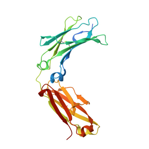
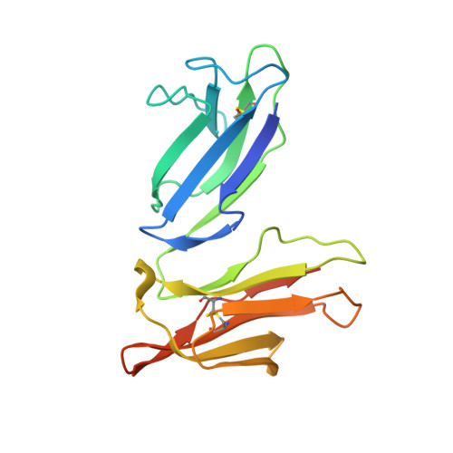
Crystal structure of IIb selective Fc variant, Fc(P238D), in complex with FcgRIIb


Experimental Data Snapshot

  • Method: X-RAY DIFFRACTION
  • Resolution: 2.60 Å
  • R-Value Free: 0.272 
  • R-Value Work: 0.230 
  • R-Value Observed: 0.232 

wwPDB Validation   3D Report Full Report


This is version 2.0 of the entry. See complete history


Literature

Engineered antibody Fc variant with selectively enhanced Fc gamma RIIb binding over both Fc gamma RIIaR131 and Fc gamma RIIaH131.

Mimoto, F.Katada, H.Kadono, S.Igawa, T.Kuramochi, T.Muraoka, M.Wada, Y.Haraya, K.Miyazaki, T.Hattori, K.

(2013) Protein Eng Des Sel 26: 589-598

  • DOI: https://doi.org/10.1093/protein/gzt022
  • Primary Citation of Related Structures:  
    3WJJ, 3WJL

  • PubMed Abstract: 

    Engaging inhibitory FcγRIIb by Fc region has been recently reported to be an attractive approach for improving the efficacy of antibody therapeutics. However, the previously reported S267E/L328F variant with enhanced binding affinity to FcγRIIb, also enhances binding affinity to FcγRIIa(R131) allotype to a similar degree because FcγRIIb and FcγRIIa(R131) are structurally similar. In this study, we applied comprehensive mutagenesis and structure-guided design based on the crystal structure of the Fc/FcγRIIb complex to identify a novel Fc variant with selectively enhanced FcγRIIb binding over both FcγRIIa(R131) and FcγRIIa(H131). This novel variant has more than 200-fold stronger binding affinity to FcγRIIb than wild-type IgG1, while binding affinity to FcγRIIa(R131) and FcγRIIa(H131) is comparable with or lower than wild-type IgG1. This selectivity was achieved by conformational change of the C(H)2 domain by mutating Pro to Asp at position 238. Fc variant with increased binding to both FcγRIIb and FcγRIIa induced platelet aggregation and activation in an immune complex form in vitro while our novel variant did not. When applied to agonistic anti-CD137 IgG1 antibody, our variant greatly enhanced the agonistic activity. Thus, the selective enhancement of FcγRIIb binding achieved by our Fc variant provides a novel tool for improving the efficacy of antibody therapeutics.


  • Organizational Affiliation

    Research Division, Chugai Pharmaceutical Co., Ltd., Gotemba, Shizuoka, Japan.


Macromolecules
Find similar proteins by:  (by identity cutoff)  |  3D Structure
Entity ID: 1
MoleculeChains Sequence LengthOrganismDetailsImage
Ig gamma-1 chain C region
A, B
230Homo sapiensMutation(s): 2 
Gene Names: IGHG1
UniProt & NIH Common Fund Data Resources
Find proteins for P01857 (Homo sapiens)
Explore P01857 
Go to UniProtKB:  P01857
PHAROS:  P01857
GTEx:  ENSG00000211896 
Entity Groups  
Sequence Clusters30% Identity50% Identity70% Identity90% Identity95% Identity100% Identity
UniProt GroupP01857
Glycosylation
Glycosylation Sites: 1Go to GlyGen: P01857-2
Sequence Annotations
Expand
  • Reference Sequence
Find similar proteins by:  (by identity cutoff)  |  3D Structure
Entity ID: 2
MoleculeChains Sequence LengthOrganismDetailsImage
Low affinity immunoglobulin gamma Fc region receptor II-b179Homo sapiensMutation(s): 0 
Gene Names: CD32FCG2FCGR2BFCGR2CIGFR2
UniProt & NIH Common Fund Data Resources
Find proteins for P31994 (Homo sapiens)
Explore P31994 
Go to UniProtKB:  P31994
PHAROS:  P31994
GTEx:  ENSG00000072694 
Entity Groups  
Sequence Clusters30% Identity50% Identity70% Identity90% Identity95% Identity100% Identity
UniProt GroupP31994
Sequence Annotations
Expand
  • Reference Sequence
Oligosaccharides

Help

Entity ID: 3
MoleculeChains Length2D Diagram Glycosylation3D Interactions
beta-D-galactopyranose-(1-4)-2-acetamido-2-deoxy-beta-D-glucopyranose-(1-2)-alpha-D-mannopyranose-(1-6)-[2-acetamido-2-deoxy-beta-D-glucopyranose-(1-2)-alpha-D-mannopyranose-(1-3)]beta-D-mannopyranose-(1-4)-2-acetamido-2-deoxy-beta-D-glucopyranose-(1-4)-[beta-L-fucopyranose-(1-6)]2-acetamido-2-deoxy-beta-D-glucopyranose
D, E
9N-Glycosylation
Glycosylation Resources
GlyTouCan:  G33244HE
GlyCosmos:  G33244HE
GlyGen:  G33244HE
Experimental Data & Validation

Experimental Data

  • Method: X-RAY DIFFRACTION
  • Resolution: 2.60 Å
  • R-Value Free: 0.272 
  • R-Value Work: 0.230 
  • R-Value Observed: 0.232 
  • Space Group: P 1 21 1
Unit Cell:
Length ( Å )Angle ( ˚ )
a = 48.85α = 90
b = 76.01β = 100.7
c = 115.08γ = 90
Software Package:
Software NamePurpose
SCALEPACKdata scaling
REFMACrefinement
PDB_EXTRACTdata extraction
ADSCdata collection
XDSdata reduction
XSCALEdata scaling
PHASERphasing

Structure Validation

View Full Validation Report


Entry History 

Deposition Data

Revision History  (Full details and data files)

  • Version 1.0: 2013-11-13
    Type: Initial release
  • Version 1.1: 2014-11-26
    Changes: Other
  • Version 1.2: 2018-06-20
    Changes: Data collection, Source and taxonomy
  • Version 2.0: 2020-07-29
    Type: Remediation
    Reason: Carbohydrate remediation
    Changes: Atomic model, Data collection, Database references, Derived calculations, Structure summary