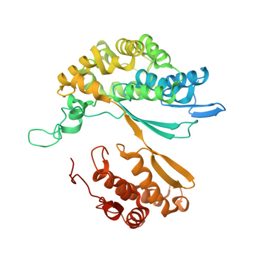
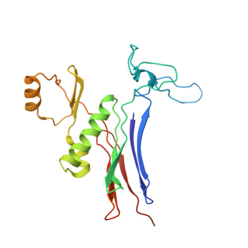
3WHR

Crystal structure of gamma-glutamyltranspeptidase from Bacillus subtilis (crystal soaked for 3min. in acivicin soln. )


Experimental Data Snapshot

  • Method: X-RAY DIFFRACTION
  • Resolution: 1.58 Å
  • R-Value Free: 0.184 
  • R-Value Work: 0.168 
  • R-Value Observed: 0.168 

Starting Model: experimental
View more details

wwPDB Validation   3D Report Full Report


This is version 1.2 of the entry. See complete history


Literature

Structure of Bacillus subtilis gamma-glutamyltranspeptidase in complex with acivicin: diversity of the binding mode of a classical and electrophilic active-site-directed glutamate analogue.

Ida, T.Suzuki, H.Fukuyama, K.Hiratake, J.Wada, K.

(2014) Acta Crystallogr D Biol Crystallogr 70: 607-614

  • DOI: https://doi.org/10.1107/S1399004713031222
  • Primary Citation of Related Structures:  
    3WHQ, 3WHR, 3WHS

  • PubMed Abstract: 

    γ-Glutamyltranspeptidase (GGT) is an enzyme that plays a central role in glutathione metabolism, and acivicin is a classical inhibitor of GGT. Here, the structure of acivicin bound to Bacillus subtilis GGT determined by X-ray crystallography to 1.8 Å resolution is presented, in which it binds to the active site in a similar manner to that in Helicobacter pylori GGT, but in a different binding mode to that in Escherichia coli GGT. In B. subtilis GGT, acivicin is bound covalently through its C3 atom with sp2 hybridization to Thr403 Oγ, the catalytic nucleophile of the enzyme. The results show that acivicin-binding sites are common, but the binding manners and orientations of its five-membered dihydroisoxazole ring are diverse in the binding pockets of GGTs.


  • Organizational Affiliation

    Department of Biological Sciences, Graduate School of Science, Osaka University, Toyonaka, Osaka 560-0043, Japan.


Macromolecules
Find similar proteins by:  (by identity cutoff)  |  3D Structure
Entity ID: 1
MoleculeChains Sequence LengthOrganismDetailsImage
Gamma-glutamyltranspeptidase large chain418Bacillus subtilis subsp. subtilis str. 168Mutation(s): 0 
Gene Names: ggtBSU18410
EC: 2.3.2.2 (PDB Primary Data), 3.4.19.13 (PDB Primary Data)
UniProt
Find proteins for P54422 (Bacillus subtilis (strain 168))
Explore P54422 
Go to UniProtKB:  P54422
Entity Groups  
Sequence Clusters30% Identity50% Identity70% Identity90% Identity95% Identity100% Identity
UniProt GroupP54422
Sequence Annotations
Expand
  • Reference Sequence
Find similar proteins by:  (by identity cutoff)  |  3D Structure
Entity ID: 2
MoleculeChains Sequence LengthOrganismDetailsImage
Gamma-glutamyltranspeptidase small chain185Bacillus subtilis subsp. subtilis str. 168Mutation(s): 0 
Gene Names: ggtBSU18410
EC: 2.3.2.2 (PDB Primary Data), 3.4.19.13 (PDB Primary Data)
UniProt
Find proteins for P54422 (Bacillus subtilis (strain 168))
Explore P54422 
Go to UniProtKB:  P54422
Entity Groups  
Sequence Clusters30% Identity50% Identity70% Identity90% Identity95% Identity100% Identity
UniProt GroupP54422
Sequence Annotations
Expand
  • Reference Sequence
Experimental Data & Validation

Experimental Data

  • Method: X-RAY DIFFRACTION
  • Resolution: 1.58 Å
  • R-Value Free: 0.184 
  • R-Value Work: 0.168 
  • R-Value Observed: 0.168 
  • Space Group: P 21 21 21
Unit Cell:
Length ( Å )Angle ( ˚ )
a = 58.786α = 90
b = 71.802β = 90
c = 142.416γ = 90
Software Package:
Software NamePurpose
BSSdata collection
MOLREPphasing
REFMACrefinement
HKL-2000data reduction
HKL-2000data scaling

Structure Validation

View Full Validation Report


Entry History 

Deposition Data

Revision History  (Full details and data files)

  • Version 1.0: 2014-02-19
    Type: Initial release
  • Version 1.1: 2019-12-25
    Changes: Database references
  • Version 1.2: 2023-11-08
    Changes: Data collection, Database references, Refinement description