3WEO

Sugar beet alpha-glucosidase with acarviosyl-maltohexaose


Experimental Data Snapshot

  • Method: X-RAY DIFFRACTION
  • Resolution: 1.45 Å
  • R-Value Free: 0.150 
  • R-Value Work: 0.123 
  • R-Value Observed: 0.124 

Starting Model: experimental
View more details

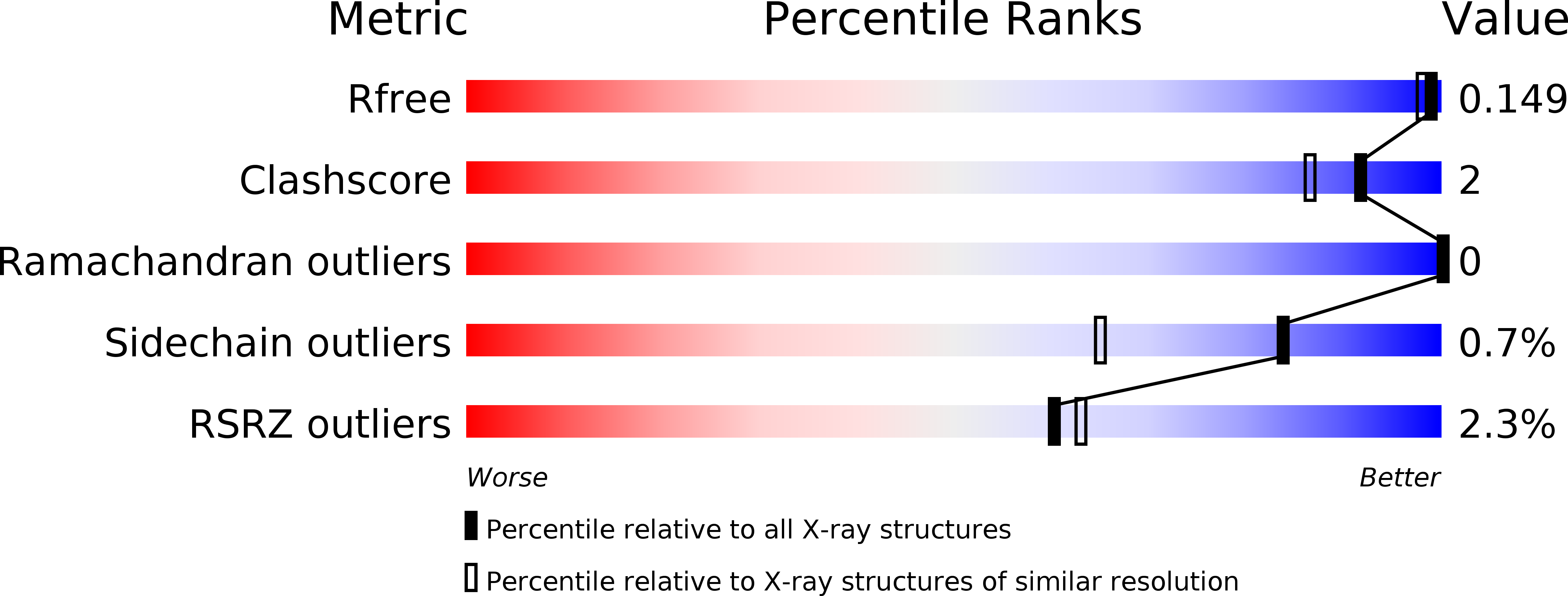
wwPDB Validation   3D Report Full Report


Ligand Structure Quality Assessment 


This is version 2.1 of the entry. See complete history


Literature

Structural advantage of sugar beet alpha-glucosidase to stabilize the Michaelis complex with long-chain substrate

Tagami, T.Yamashita, K.Okuyama, M.Mori, H.Yao, M.Kimura, A.

(2014) J Biol Chem 290: 1796-1803

  • DOI: https://doi.org/10.1074/jbc.M114.606939
  • Primary Citation of Related Structures:  
    3WEL, 3WEM, 3WEN, 3WEO

  • PubMed Abstract: 

    The α-glucosidase from sugar beet (SBG) is an exo-type glycosidase. The enzyme has a pocket-shaped active site, but efficiently hydrolyzes longer maltooligosaccharides and soluble starch due to lower Km and higher kcat/Km for such substrates. To obtain structural insights into the mechanism governing its unique substrate specificity, a series of acarviosyl-maltooligosaccharides was employed for steady-state kinetic and structural analyses. The acarviosyl-maltooligosaccharides have a longer maltooligosaccharide moiety compared with the maltose moiety of acarbose, which is known to be the transition state analog of α-glycosidases. The clear correlation obtained between log Ki of the acarviosyl-maltooligosaccharides and log(Km/kcat) for hydrolysis of maltooligosaccharides suggests that the acarviosyl-maltooligosaccharides are transition state mimics. The crystal structure of the enzyme bound with acarviosyl-maltohexaose reveals that substrate binding at a distance from the active site is maintained largely by van der Waals interactions, with the four glucose residues at the reducing terminus of acarviosyl-maltohexaose retaining a left-handed single-helical conformation, as also observed in cycloamyloses and single helical V-amyloses. The kinetic behavior and structural features suggest that the subsite structure suitable for the stable conformation of amylose lowers the Km for long-chain substrates, which in turn is responsible for higher specificity of the longer substrates.


  • Organizational Affiliation

    From the Research Faculty of Agriculture.


Macromolecules
Find similar proteins by:  (by identity cutoff)  |  3D Structure
Entity ID: 1
MoleculeChains Sequence LengthOrganismDetailsImage
Alpha-glucosidase913Beta vulgarisMutation(s): 0 
EC: 3.2.1.20
UniProt
Find proteins for L0N7E5 (Beta vulgaris)
Explore L0N7E5 
Go to UniProtKB:  L0N7E5
Entity Groups  
Sequence Clusters30% Identity50% Identity70% Identity90% Identity95% Identity100% Identity
UniProt GroupL0N7E5
Glycosylation
Glycosylation Sites: 3
Sequence Annotations
Expand
  • Reference Sequence
Oligosaccharides

Help

Entity ID: 2
MoleculeChains Length2D Diagram Glycosylation3D Interactions
alpha-L-fucopyranose-(1-3)-[2-acetamido-2-deoxy-beta-D-glucopyranose-(1-4)]2-acetamido-2-deoxy-beta-D-glucopyranose
B
3N-Glycosylation
Glycosylation Resources
GlyTouCan:  G11971MR
GlyCosmos:  G11971MR
GlyGen:  G11971MR
Entity ID: 3
MoleculeChains Length2D Diagram Glycosylation3D Interactions
2-acetamido-2-deoxy-beta-D-glucopyranose-(1-4)-2-acetamido-2-deoxy-beta-D-glucopyranose
C
2N-Glycosylation
Glycosylation Resources
GlyTouCan:  G42666HT
GlyCosmos:  G42666HT
GlyGen:  G42666HT
Entity ID: 4
MoleculeChains Length2D Diagram Glycosylation3D Interactions
4,6-dideoxy-4-{[(1S,4R,5S,6S)-4,5,6-trihydroxy-3-(hydroxymethyl)cyclohex-2-en-1-yl]amino}-alpha-D-glucopyranose-(1-4)-alpha-D-glucopyranose-(1-4)-alpha-D-glucopyranose-(1-4)-alpha-D-glucopyranose-(1-4)-alpha-D-glucopyranose-(1-4)-alpha-D-glucopyranose-(1-4)-alpha-D-glucopyranose
D
7N/AN/A
Glycosylation Resources
GlyTouCan:  G67262XF
GlyCosmos:  G67262XF
Experimental Data & Validation

Experimental Data

  • Method: X-RAY DIFFRACTION
  • Resolution: 1.45 Å
  • R-Value Free: 0.150 
  • R-Value Work: 0.123 
  • R-Value Observed: 0.124 
  • Space Group: P 21 21 21
Unit Cell:
Length ( Å )Angle ( ˚ )
a = 86.166α = 90
b = 99.154β = 90
c = 107.354γ = 90
Software Package:
Software NamePurpose
XSCALEdata scaling
PHENIXrefinement
PDB_EXTRACTdata extraction
BSSdata collection
XDSdata reduction
XDSdata scaling
PHENIXphasing

Structure Validation

View Full Validation Report


Ligand Structure Quality Assessment 


Entry History 

Deposition Data

Revision History  (Full details and data files)

  • Version 1.0: 2014-07-16
    Type: Initial release
  • Version 1.1: 2014-07-30
    Changes: Other
  • Version 1.2: 2014-12-31
    Changes: Database references
  • Version 1.3: 2015-02-04
    Changes: Database references
  • Version 2.0: 2020-07-29
    Type: Remediation
    Reason: Carbohydrate remediation
    Changes: Advisory, Atomic model, Data collection, Derived calculations, Non-polymer description, Structure summary
  • Version 2.1: 2023-11-08
    Changes: Data collection, Database references, Refinement description, Structure summary