3WDL

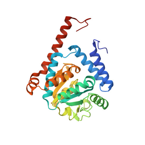
Crystal structure of 4-phosphopantoate-beta-alanine ligase complexed with ATP


Experimental Data Snapshot

  • Method: X-RAY DIFFRACTION
  • Resolution: 2.40 Å
  • R-Value Free: 0.282 
  • R-Value Work: 0.227 
  • R-Value Observed: 0.227 

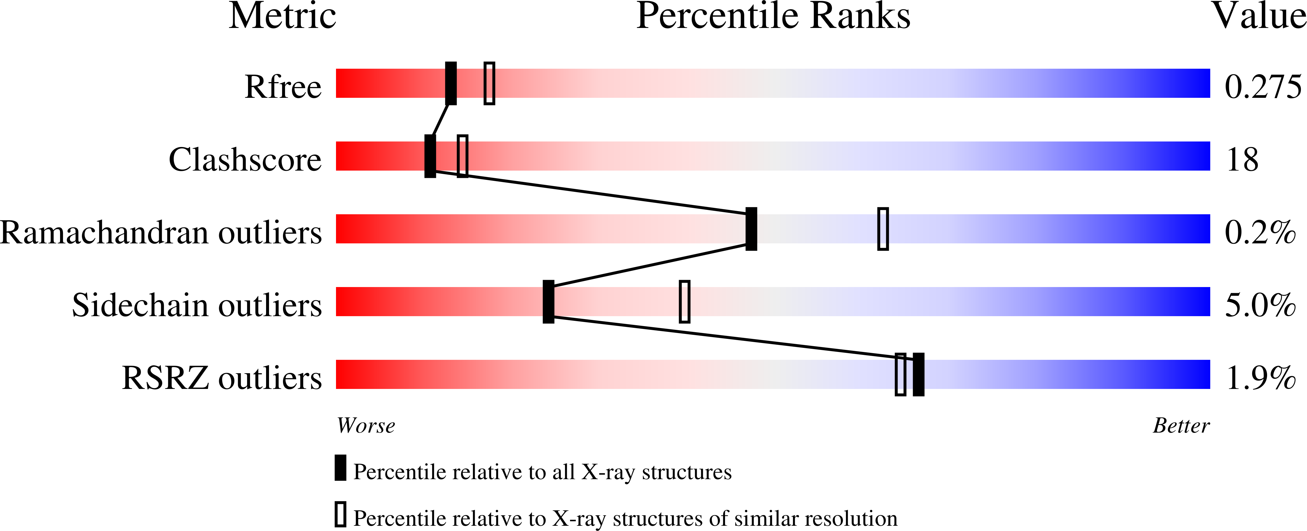
wwPDB Validation   3D Report Full Report


Ligand Structure Quality Assessment 


This is version 1.2 of the entry. See complete history


Literature

Crystal structure of phosphopantothenate synthetase from Thermococcus kodakarensis

Kishimoto, A.Kita, A.Ishibashi, T.Tomita, H.Yokooji, Y.Imanaka, T.Atomi, H.Miki, K.

(2014) Proteins 82: 1924-1936

  • DOI: https://doi.org/10.1002/prot.24546
  • Primary Citation of Related Structures:  
    3WDK, 3WDL, 3WDM

  • PubMed Abstract: 

    Bacteria/eukaryotes share a common pathway for coenzyme A biosynthesis which involves two enzymes to convert pantoate to 4'-phosphopantothenate. These two enzymes are absent in almost all archaea. Recently, it was reported that two novel enzymes, pantoate kinase, and phosphopantothenate synthetase (PPS), are responsible for this conversion in archaea. Here, we report the crystal structure of PPS from the hyperthermophilic archaeon, Thermococcus kodakarensis and its complexes with substrates, ATP, and ATP and 4-phosphopantoate. PPS forms an asymmetric homodimer, in which two monomers composing a dimer, deviated from the exact twofold symmetry, displaying 4°-13° distortion. The structural features are consistent with the mutagenesis data and the results of biochemical experiments previously reported. Based on these structures, we discuss the catalytic mechanism by which PPS produces phosphopantoyl adenylate, which is thought to be a reaction intermediate.


  • Organizational Affiliation

    Department of Chemistry, Graduate School of Science, Kyoto University, Sakyo-ku, Kyoto, 606-8502, Japan.


Macromolecules
Find similar proteins by:  (by identity cutoff)  |  3D Structure
Entity ID: 1
MoleculeChains Sequence LengthOrganismDetailsImage
4-phosphopantoate--beta-alanine ligase
A, B, C, D
261Thermococcus kodakarensis KOD1Mutation(s): 0 
Gene Names: TK1686
EC: 6.3.2.36
UniProt
Find proteins for Q5JIZ8 (Thermococcus kodakarensis (strain ATCC BAA-918 / JCM 12380 / KOD1))
Explore Q5JIZ8 
Go to UniProtKB:  Q5JIZ8
Entity Groups  
Sequence Clusters30% Identity50% Identity70% Identity90% Identity95% Identity100% Identity
UniProt GroupQ5JIZ8
Sequence Annotations
Expand
  • Reference Sequence
Experimental Data & Validation

Experimental Data

  • Method: X-RAY DIFFRACTION
  • Resolution: 2.40 Å
  • R-Value Free: 0.282 
  • R-Value Work: 0.227 
  • R-Value Observed: 0.227 
  • Space Group: P 1 21 1
Unit Cell:
Length ( Å )Angle ( ˚ )
a = 79.655α = 90
b = 80.071β = 110.43
c = 83.998γ = 90
Software Package:
Software NamePurpose
HKL-2000data collection
MOLREPphasing
CNSrefinement
HKL-2000data reduction
HKL-2000data scaling

Structure Validation

View Full Validation Report


Ligand Structure Quality Assessment 


Entry History 

Deposition Data

Revision History  (Full details and data files)

  • Version 1.0: 2014-04-02
    Type: Initial release
  • Version 1.1: 2017-02-22
    Changes: Database references
  • Version 1.2: 2024-03-20
    Changes: Data collection, Database references, Derived calculations